Sandbox Reserved 1791
From Proteopedia
(Difference between revisions)
| Line 11: | Line 11: | ||
=== Transmembrane Region=== | === Transmembrane Region=== | ||
<scene name='95/952720/Transmembrane_region_spin/5'>The Transmembrane Region</scene> (<scene name='95/952720/Transmembrane_region_top-view/2'>top-view</scene>) is embedded within the cell membrane. Like other G-protein receptors, it is made up of a 7-pass helix <ref name="Faust"> DOI 10.1038/s41586-022-05159-1</ref>. It is made up of about 284 residues. The transmembrane region is surrounded by a "belt" of <scene name='95/952719/Tmd_cholesterol_spin/2'>15 cholesterols</scene>. When cholesterol binding sites are mutated such that they are unfunctional, TSHR activity decreases. Thus, the cholesterols are important for TSHR function <ref name="Duan"> DOI 10.1038/s41586-022-05173-3</ref>. Additionally, at the N-terminus, the transmembrane region binds to the <scene name='95/952720/Transmembrane_region_spin/4'>G-proteins</scene>, which are located intracellularly. | <scene name='95/952720/Transmembrane_region_spin/5'>The Transmembrane Region</scene> (<scene name='95/952720/Transmembrane_region_top-view/2'>top-view</scene>) is embedded within the cell membrane. Like other G-protein receptors, it is made up of a 7-pass helix <ref name="Faust"> DOI 10.1038/s41586-022-05159-1</ref>. It is made up of about 284 residues. The transmembrane region is surrounded by a "belt" of <scene name='95/952719/Tmd_cholesterol_spin/2'>15 cholesterols</scene>. When cholesterol binding sites are mutated such that they are unfunctional, TSHR activity decreases. Thus, the cholesterols are important for TSHR function <ref name="Duan"> DOI 10.1038/s41586-022-05173-3</ref>. Additionally, at the N-terminus, the transmembrane region binds to the <scene name='95/952720/Transmembrane_region_spin/4'>G-proteins</scene>, which are located intracellularly. | ||
| - | |||
| - | |||
=== Leucine Rich Domain=== | === Leucine Rich Domain=== | ||
| - | |||
The <scene name='95/952719/Lrrd/1'>Leucine Rich Repeat Domain (LRRD)</scene> is part of the extracellular region of TSHR. It is made up of about 280 different residues. Connected to its C-terminus is the Hinge Region. It is made up of an extensive parallel β-sheet. This β-sheet is where TSH binds and is called the binding pocket<ref name="Duan"> DOI 10.1038/s41586-022-05173-3</ref>. The <scene name='95/952719/Binding_pocket/4'>binding pocket</scene> is a concave structure with many polar residues. This pocket is where the TSH antibody and agonist K1 bind as well as the agonist M22. These structures interact with specific residues to result in a structural change of the molecule. There is a mutation done by N-glycans at asparagine residues that plays a large role in the binding of TSH. The negative charge on these glycans contributes to the polarity of the binding pocket which mediates the binding efficiency of TSH. It has been shown that four of the five N glycan sites must be glycosylated to be in the active form<ref name="Fokina">Fokina, E.F., Shpakov, A.O. Thyroid-Stimulating Hormone Receptor: the Role in the Development of Thyroid Pathology and Its Correction. J Evol Biochem Phys 58, 1439–1454 (2022). [DOI:10.1134/S0022093022050143 https://doi.org/10.1134/S0022093022050143]</ref>. | The <scene name='95/952719/Lrrd/1'>Leucine Rich Repeat Domain (LRRD)</scene> is part of the extracellular region of TSHR. It is made up of about 280 different residues. Connected to its C-terminus is the Hinge Region. It is made up of an extensive parallel β-sheet. This β-sheet is where TSH binds and is called the binding pocket<ref name="Duan"> DOI 10.1038/s41586-022-05173-3</ref>. The <scene name='95/952719/Binding_pocket/4'>binding pocket</scene> is a concave structure with many polar residues. This pocket is where the TSH antibody and agonist K1 bind as well as the agonist M22. These structures interact with specific residues to result in a structural change of the molecule. There is a mutation done by N-glycans at asparagine residues that plays a large role in the binding of TSH. The negative charge on these glycans contributes to the polarity of the binding pocket which mediates the binding efficiency of TSH. It has been shown that four of the five N glycan sites must be glycosylated to be in the active form<ref name="Fokina">Fokina, E.F., Shpakov, A.O. Thyroid-Stimulating Hormone Receptor: the Role in the Development of Thyroid Pathology and Its Correction. J Evol Biochem Phys 58, 1439–1454 (2022). [DOI:10.1134/S0022093022050143 https://doi.org/10.1134/S0022093022050143]</ref>. | ||
| - | |||
=== Hinge Region=== | === Hinge Region=== | ||
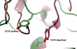
The <scene name='95/952719/Hinge_region_spin/3'>Higne Region</scene>(purple-blue) connects the Transmembrane Region to the Leucine Rich Domain. It is sometimes referred to as the signaling specificity domain because there is some evidence suggesting that this region is important in both TSH binding and signal transduction. <ref name="Chen">Chen CR, McLachlan SM, Rapoport B. Thyrotropin (TSH) receptor residue E251 in the extracellular leucine-rich repeat domain is critical for linking TSH binding to receptor activation. Endocrinology. 2010 Apr;151(4):1940-7. doi: 10.1210/en.2009-1430. Epub 2010 Feb 24. PMID: 20181794; PMCID: PMC2851189. [DOI 10.1210/en.2009-1430 https://www.ncbi.nlm.nih.gov/pmc/articles/PMC2851189/]</ref>. It is made up of two α-helices that are connected via disulfide bonds(shown in yellow). Interactions between these two helices and TSH help orient TSH properly. These interactions are essential for TSH binding, however, they are not required for the activation of TSHR. Conformational changes in this region, specifically the orientation of <scene name='95/952719/Hinge_region_residues/3'>Y279</scene>, are responsible for bringing TSHR into the active state <ref name="Faust"/>. | The <scene name='95/952719/Hinge_region_spin/3'>Higne Region</scene>(purple-blue) connects the Transmembrane Region to the Leucine Rich Domain. It is sometimes referred to as the signaling specificity domain because there is some evidence suggesting that this region is important in both TSH binding and signal transduction. <ref name="Chen">Chen CR, McLachlan SM, Rapoport B. Thyrotropin (TSH) receptor residue E251 in the extracellular leucine-rich repeat domain is critical for linking TSH binding to receptor activation. Endocrinology. 2010 Apr;151(4):1940-7. doi: 10.1210/en.2009-1430. Epub 2010 Feb 24. PMID: 20181794; PMCID: PMC2851189. [DOI 10.1210/en.2009-1430 https://www.ncbi.nlm.nih.gov/pmc/articles/PMC2851189/]</ref>. It is made up of two α-helices that are connected via disulfide bonds(shown in yellow). Interactions between these two helices and TSH help orient TSH properly. These interactions are essential for TSH binding, however, they are not required for the activation of TSHR. Conformational changes in this region, specifically the orientation of <scene name='95/952719/Hinge_region_residues/3'>Y279</scene>, are responsible for bringing TSHR into the active state <ref name="Faust"/>. | ||
| Line 29: | Line 25: | ||
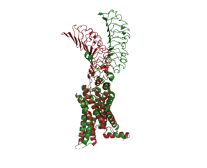
[[Image:Image-Inactive Inactive Proteopedia.png|300 px|right|thumb| Figure 2: An overview of the Inactive (red) vs Active (green) state of TSHR. PDB: 7WX5]] | [[Image:Image-Inactive Inactive Proteopedia.png|300 px|right|thumb| Figure 2: An overview of the Inactive (red) vs Active (green) state of TSHR. PDB: 7WX5]] | ||
[[Image:Inactive v active residue.png|300 px|right|thumb| Figure 3: A zoomed in view of the Y279 residue in the Hinge Region of TSHR, showing the 6 angstrom move of Y279 during the activation of TSHR. Active TSHR is shown in green (PDB: 7t9i) and inactive TSHR is shown in pink (PDB: 7t9m).]] | [[Image:Inactive v active residue.png|300 px|right|thumb| Figure 3: A zoomed in view of the Y279 residue in the Hinge Region of TSHR, showing the 6 angstrom move of Y279 during the activation of TSHR. Active TSHR is shown in green (PDB: 7t9i) and inactive TSHR is shown in pink (PDB: 7t9m).]] | ||
| - | + | In its resting state without TSH, TSHR is in the <scene name='95/952720/Inactivetshr/7'>inactive state</scene>, also considered the "down" state because the LRRD is pointing down. When TSH binds to TSHR, steric clashes between TSH and the cell-membrane cause TSHR to take on the <scene name='95/952720/Inactivetshr/6'>active or "up" state</scene>. During this transition, the extracellular LRD rotates 55° along an axis making it perpendicular to the cell membrane. This rotation is initiated by conformational changes within the <scene name='95/952720/Hinge_region_spin/3'>Hinge Region</scene>, specifically at the <scene name='95/952720/Hinge_region_residues/2'>TYR279 residue</scene>. Y279 moves 6 Å relative to I486, a residue located in the Transmembrane Region <ref name="Faust"/>. The active form is favored when <scene name='95/952719/Active_form/4'>TSHR is bound to TSH or M22</scene>. The structure can be seen as straight. The inactive form is found when <scene name='95/952719/Inactive_form/6'>TSHR is bound with K1</scene> of TSHR is found when bound with K1. The overall structure of the molecule is bent when K1 is bound. | |
== Specific Residues and Interactions== | == Specific Residues and Interactions== | ||
Revision as of 17:51, 14 April 2023
| This Sandbox is Reserved from February 27 through August 31, 2023 for use in the course CH462 Biochemistry II taught by R. Jeremy Johnson at the Butler University, Indianapolis, USA. This reservation includes Sandbox Reserved 1765 through Sandbox Reserved 1795. |
To get started:
More help: Help:Editing |
Thyroid Stimulating Hormone Receptor (TSHR)
| |||||||||||
References
- ↑ 1.0 1.1 1.2 Faust B, Billesbolle CB, Suomivuori CM, Singh I, Zhang K, Hoppe N, Pinto AFM, Diedrich JK, Muftuoglu Y, Szkudlinski MW, Saghatelian A, Dror RO, Cheng Y, Manglik A. Autoantibody mimicry of hormone action at the thyrotropin receptor. Nature. 2022 Aug 8. pii: 10.1038/s41586-022-05159-1. doi:, 10.1038/s41586-022-05159-1. PMID:35940205 doi:http://dx.doi.org/10.1038/s41586-022-05159-1
- ↑ 2.0 2.1 Duan J, Xu P, Luan X, Ji Y, He X, Song N, Yuan Q, Jin Y, Cheng X, Jiang H, Zheng J, Zhang S, Jiang Y, Xu HE. Hormone- and antibody-mediated activation of the thyrotropin receptor. Nature. 2022 Aug 8. pii: 10.1038/s41586-022-05173-3. doi:, 10.1038/s41586-022-05173-3. PMID:35940204 doi:http://dx.doi.org/10.1038/s41586-022-05173-3
- ↑ Fokina, E.F., Shpakov, A.O. Thyroid-Stimulating Hormone Receptor: the Role in the Development of Thyroid Pathology and Its Correction. J Evol Biochem Phys 58, 1439–1454 (2022). [DOI:10.1134/S0022093022050143 https://doi.org/10.1134/S0022093022050143]
- ↑ Chen CR, McLachlan SM, Rapoport B. Thyrotropin (TSH) receptor residue E251 in the extracellular leucine-rich repeat domain is critical for linking TSH binding to receptor activation. Endocrinology. 2010 Apr;151(4):1940-7. doi: 10.1210/en.2009-1430. Epub 2010 Feb 24. PMID: 20181794; PMCID: PMC2851189. [DOI 10.1210/en.2009-1430 https://www.ncbi.nlm.nih.gov/pmc/articles/PMC2851189/]
- ↑ Nunez Miguel R, Sanders J, Chirgadze DY, Furmaniak J, Rees Smith B. Thyroid stimulating autoantibody M22 mimics TSH binding to the TSH receptor leucine rich domain: a comparative structural study of protein-protein interactions. J Mol Endocrinol. 2009 May;42(5):381-95. Epub 2009 Feb 16. PMID:19221175 doi:10.1677/JME-08-0152
- ↑ Smits G, Govaerts C, Nubourgh I, Pardo L, Vassart G, Costagliola S. Lysine 183 and glutamic acid 157 of the TSH receptor: two interacting residues with a key role in determining specificity toward TSH and human CG. Mol Endocrinol. 2002 Apr;16(4):722-35. doi: 10.1210/mend.16.4.0815. PMID: 11923469. [DOI: 10.1210/mend.16.4.0815 https://pubmed.ncbi.nlm.nih.gov/11923469/]
- ↑ 7.0 7.1 Chiovato L, Magri F, Carlé A. Hypothyroidism in Context: Where We've Been and Where We're Going. Adv Ther. 2019 Sep;36(Suppl 2):47-58. doi: 10.1007/s12325-019-01080-8. Epub 2019 Sep 4. PMID: 31485975; PMCID: PMC6822815. [DOI: 10.1007/s12325-019-01080-8 https://pubmed.ncbi.nlm.nih.gov/31485975/]
![Figure 1: An overview of the Thyroid System. A depiction of signaling cascade from the hypothalamus ending in the release of TSH causing T3 and T4 production and its effects. The mechanism of regulation also shown by negative feedback from the T3 and T4 hormones. Source: [1]](/wiki/images/thumb/1/1d/TSH_system1.png/300px-TSH_system1.png)


![Figure 4: T3 and T4 role in TSH concentration: Highlighting the problem when under or overactive on the metabolism. When an antibody is bound to TSHR and cannot respond to the negative feedback look the metabolism experiences a shift outside of equilibrium resulting in a wide array of side effects. [2]](/wiki/images/thumb/5/5f/T3t4levels.jpeg/400px-T3t4levels.jpeg)
