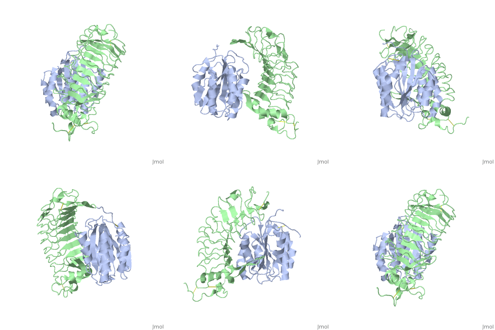
|
|
| Line 3: |
Line 3: |
| | <StructureSection load='1m10' size='340' side='right'caption='[[1m10]], [[Resolution|resolution]] 3.10Å' scene=''> | | <StructureSection load='1m10' size='340' side='right'caption='[[1m10]], [[Resolution|resolution]] 3.10Å' scene=''> |
| | == Structural highlights == | | == Structural highlights == |
| - | <table><tr><td colspan='2'>[[1m10]] is a 2 chain structure with sequence from [https://en.wikipedia.org/wiki/Human Human]. Full crystallographic information is available from [http://oca.weizmann.ac.il/oca-bin/ocashort?id=1M10 OCA]. For a <b>guided tour on the structure components</b> use [https://proteopedia.org/fgij/fg.htm?mol=1M10 FirstGlance]. <br> | + | <table><tr><td colspan='2'>[[1m10]] is a 2 chain structure with sequence from [https://en.wikipedia.org/wiki/Homo_sapiens Homo sapiens]. Full crystallographic information is available from [http://oca.weizmann.ac.il/oca-bin/ocashort?id=1M10 OCA]. For a <b>guided tour on the structure components</b> use [https://proteopedia.org/fgij/fg.htm?mol=1M10 FirstGlance]. <br> |
| - | </td></tr><tr id='related'><td class="sblockLbl"><b>[[Related_structure|Related:]]</b></td><td class="sblockDat"><div style='overflow: auto; max-height: 3em;'>[[1m0z|1m0z]]</div></td></tr> | + | </td></tr><tr id='method'><td class="sblockLbl"><b>[[Empirical_models|Method:]]</b></td><td class="sblockDat" id="methodDat">X-ray diffraction, [[Resolution|Resolution]] 3.1Å</td></tr> |
| - | <tr id='gene'><td class="sblockLbl"><b>[[Gene|Gene:]]</b></td><td class="sblockDat">VWF ([https://www.ncbi.nlm.nih.gov/Taxonomy/Browser/wwwtax.cgi?mode=Info&srchmode=5&id=9606 HUMAN]), GP1BA ([https://www.ncbi.nlm.nih.gov/Taxonomy/Browser/wwwtax.cgi?mode=Info&srchmode=5&id=9606 HUMAN])</td></tr>
| + | |
| | <tr id='resources'><td class="sblockLbl"><b>Resources:</b></td><td class="sblockDat"><span class='plainlinks'>[https://proteopedia.org/fgij/fg.htm?mol=1m10 FirstGlance], [http://oca.weizmann.ac.il/oca-bin/ocaids?id=1m10 OCA], [https://pdbe.org/1m10 PDBe], [https://www.rcsb.org/pdb/explore.do?structureId=1m10 RCSB], [https://www.ebi.ac.uk/pdbsum/1m10 PDBsum], [https://prosat.h-its.org/prosat/prosatexe?pdbcode=1m10 ProSAT]</span></td></tr> | | <tr id='resources'><td class="sblockLbl"><b>Resources:</b></td><td class="sblockDat"><span class='plainlinks'>[https://proteopedia.org/fgij/fg.htm?mol=1m10 FirstGlance], [http://oca.weizmann.ac.il/oca-bin/ocaids?id=1m10 OCA], [https://pdbe.org/1m10 PDBe], [https://www.rcsb.org/pdb/explore.do?structureId=1m10 RCSB], [https://www.ebi.ac.uk/pdbsum/1m10 PDBsum], [https://prosat.h-its.org/prosat/prosatexe?pdbcode=1m10 ProSAT]</span></td></tr> |
| | </table> | | </table> |
| | == Disease == | | == Disease == |
| - | [[https://www.uniprot.org/uniprot/VWF_HUMAN VWF_HUMAN]] Defects in VWF are the cause of von Willebrand disease type 1 (VWD1) [MIM:[https://omim.org/entry/193400 193400]]. A common hemorrhagic disorder due to defects in von Willebrand factor protein and resulting in impaired platelet aggregation. Von Willebrand disease type 1 is characterized by partial quantitative deficiency of circulating von Willebrand factor, that is otherwise structurally and functionally normal. Clinical manifestations are mucocutaneous bleeding, such as epistaxis and menorrhagia, and prolonged bleeding after surgery or trauma.<ref>PMID:10887119</ref> <ref>PMID:11698279</ref> Defects in VWF are the cause of von Willebrand disease type 2 (VWD2) [MIM:[https://omim.org/entry/613554 613554]]. A hemorrhagic disorder due to defects in von Willebrand factor protein and resulting in impaired platelet aggregation. Von Willebrand disease type 2 is characterized by qualitative deficiency and functional anomalies of von Willebrand factor. It is divided in different subtypes including 2A, 2B, 2M and 2N (Normandy variant). The mutant VWF protein in types 2A, 2B and 2M are defective in their platelet-dependent function, whereas the mutant protein in type 2N is defective in its ability to bind factor VIII. Clinical manifestations are mucocutaneous bleeding, such as epistaxis and menorrhagia, and prolonged bleeding after surgery or trauma. Defects in VWF are the cause of von Willebrand disease type 3 (VWD3) [MIM:[https://omim.org/entry/277480 277480]]. A severe hemorrhagic disorder due to a total or near total absence of von Willebrand factor in the plasma and cellular compartments, also leading to a profound deficiency of plasmatic factor VIII. Bleeding usually starts in infancy and can include epistaxis, recurrent mucocutaneous bleeding, excessive bleeding after minor trauma, and hemarthroses. [[https://www.uniprot.org/uniprot/GP1BA_HUMAN GP1BA_HUMAN]] Genetic variations in GP1BA may be a cause of susceptibility to non-arteritic anterior ischemic optic neuropathy (NAION) [MIM:[https://omim.org/entry/258660 258660]]. NAION is an ocular disease due to ischemic injury to the optic nerve. It usually affects the optic disk and leads to visual loss and optic disk swelling of a pallid nature. Visual loss is usually sudden, or over a few days at most and is usually permanent, with some recovery possibly occurring within the first weeks or months. Patients with small disks having smaller or non-existent cups have an anatomical predisposition for non-arteritic anterior ischemic optic neuropathy. As an ischemic episode evolves, the swelling compromises circulation, with a spiral of ischemia resulting in further neuronal damage.<ref>PMID:14711733</ref> Defects in GP1BA are a cause of Bernard-Soulier syndrome (BSS) [MIM:[https://omim.org/entry/231200 231200]]; also known as giant platelet disease (GPD). BSS patients have unusually large platelets and have a clinical bleeding tendency.<ref>PMID:1730088</ref> <ref>PMID:7690774</ref> <ref>PMID:7819107</ref> <ref>PMID:7873390</ref> <ref>PMID:9639514</ref> <ref>PMID:10089893</ref> Defects in GP1BA are the cause of benign mediterranean macrothrombocytopenia (BMM) [MIM:[https://omim.org/entry/153670 153670]]; also known as autosomal dominant benign Bernard-Soulier syndrome. BMM is characterized by mild or no clinical symptoms, normal platelet function, and normal megakaryocyte count.<ref>PMID:11222377</ref> Defects in GP1BA are the cause of pseudo-von Willebrand disease (VWDP) [MIM:[https://omim.org/entry/177820 177820]]. A bleeding disorder is caused by an increased affinity of GP-Ib for soluble vWF resulting in impaired hemostatic function due to the removal of vWF from the circulation.<ref>PMID:14521605</ref> <ref>PMID:2052556</ref> <ref>PMID:8486780</ref> <ref>PMID:8384898</ref>
| + | [https://www.uniprot.org/uniprot/VWF_HUMAN VWF_HUMAN] Defects in VWF are the cause of von Willebrand disease type 1 (VWD1) [MIM:[https://omim.org/entry/193400 193400]. A common hemorrhagic disorder due to defects in von Willebrand factor protein and resulting in impaired platelet aggregation. Von Willebrand disease type 1 is characterized by partial quantitative deficiency of circulating von Willebrand factor, that is otherwise structurally and functionally normal. Clinical manifestations are mucocutaneous bleeding, such as epistaxis and menorrhagia, and prolonged bleeding after surgery or trauma.<ref>PMID:10887119</ref> <ref>PMID:11698279</ref> Defects in VWF are the cause of von Willebrand disease type 2 (VWD2) [MIM:[https://omim.org/entry/613554 613554]. A hemorrhagic disorder due to defects in von Willebrand factor protein and resulting in impaired platelet aggregation. Von Willebrand disease type 2 is characterized by qualitative deficiency and functional anomalies of von Willebrand factor. It is divided in different subtypes including 2A, 2B, 2M and 2N (Normandy variant). The mutant VWF protein in types 2A, 2B and 2M are defective in their platelet-dependent function, whereas the mutant protein in type 2N is defective in its ability to bind factor VIII. Clinical manifestations are mucocutaneous bleeding, such as epistaxis and menorrhagia, and prolonged bleeding after surgery or trauma. Defects in VWF are the cause of von Willebrand disease type 3 (VWD3) [MIM:[https://omim.org/entry/277480 277480]. A severe hemorrhagic disorder due to a total or near total absence of von Willebrand factor in the plasma and cellular compartments, also leading to a profound deficiency of plasmatic factor VIII. Bleeding usually starts in infancy and can include epistaxis, recurrent mucocutaneous bleeding, excessive bleeding after minor trauma, and hemarthroses. |
| | == Function == | | == Function == |
| - | [[https://www.uniprot.org/uniprot/VWF_HUMAN VWF_HUMAN]] Important in the maintenance of hemostasis, it promotes adhesion of platelets to the sites of vascular injury by forming a molecular bridge between sub-endothelial collagen matrix and platelet-surface receptor complex GPIb-IX-V. Also acts as a chaperone for coagulation factor VIII, delivering it to the site of injury, stabilizing its heterodimeric structure and protecting it from premature clearance from plasma. [[https://www.uniprot.org/uniprot/GP1BA_HUMAN GP1BA_HUMAN]] GP-Ib, a surface membrane protein of platelets, participates in the formation of platelet plugs by binding to the A1 domain of vWF, which is already bound to the subendothelium.
| + | [https://www.uniprot.org/uniprot/VWF_HUMAN VWF_HUMAN] Important in the maintenance of hemostasis, it promotes adhesion of platelets to the sites of vascular injury by forming a molecular bridge between sub-endothelial collagen matrix and platelet-surface receptor complex GPIb-IX-V. Also acts as a chaperone for coagulation factor VIII, delivering it to the site of injury, stabilizing its heterodimeric structure and protecting it from premature clearance from plasma. |
| | == Evolutionary Conservation == | | == Evolutionary Conservation == |
| | [[Image:Consurf_key_small.gif|200px|right]] | | [[Image:Consurf_key_small.gif|200px|right]] |
| Line 38: |
Line 37: |
| | __TOC__ | | __TOC__ |
| | </StructureSection> | | </StructureSection> |
| - | [[Category: Human]] | + | [[Category: Homo sapiens]] |
| | [[Category: Large Structures]] | | [[Category: Large Structures]] |
| - | [[Category: Groot, P G.de]]
| + | [[Category: Gros P]] |
| - | [[Category: Gros, P]] | + | [[Category: Huizinga EG]] |
| - | [[Category: Huizinga, E G]] | + | [[Category: Romijn RAP]] |
| - | [[Category: Romijn, R A.P]] | + | [[Category: Schiphorst ME]] |
| - | [[Category: Schiphorst, M E]] | + | [[Category: Sixma JJ]] |
| - | [[Category: Sixma, J J]] | + | [[Category: Tsuji S]] |
| - | [[Category: Tsuji, S]] | + | [[Category: De Groot PG]] |
| - | [[Category: Blood clotting]] | + | |
| - | [[Category: Dinucleotide binding fold]]
| + | |
| - | [[Category: Hemostasis]]
| + | |
| - | [[Category: Leucine-rich repeat]]
| + | |
| Structural highlights
Disease
VWF_HUMAN Defects in VWF are the cause of von Willebrand disease type 1 (VWD1) [MIM:193400. A common hemorrhagic disorder due to defects in von Willebrand factor protein and resulting in impaired platelet aggregation. Von Willebrand disease type 1 is characterized by partial quantitative deficiency of circulating von Willebrand factor, that is otherwise structurally and functionally normal. Clinical manifestations are mucocutaneous bleeding, such as epistaxis and menorrhagia, and prolonged bleeding after surgery or trauma.[1] [2] Defects in VWF are the cause of von Willebrand disease type 2 (VWD2) [MIM:613554. A hemorrhagic disorder due to defects in von Willebrand factor protein and resulting in impaired platelet aggregation. Von Willebrand disease type 2 is characterized by qualitative deficiency and functional anomalies of von Willebrand factor. It is divided in different subtypes including 2A, 2B, 2M and 2N (Normandy variant). The mutant VWF protein in types 2A, 2B and 2M are defective in their platelet-dependent function, whereas the mutant protein in type 2N is defective in its ability to bind factor VIII. Clinical manifestations are mucocutaneous bleeding, such as epistaxis and menorrhagia, and prolonged bleeding after surgery or trauma. Defects in VWF are the cause of von Willebrand disease type 3 (VWD3) [MIM:277480. A severe hemorrhagic disorder due to a total or near total absence of von Willebrand factor in the plasma and cellular compartments, also leading to a profound deficiency of plasmatic factor VIII. Bleeding usually starts in infancy and can include epistaxis, recurrent mucocutaneous bleeding, excessive bleeding after minor trauma, and hemarthroses.
Function
VWF_HUMAN Important in the maintenance of hemostasis, it promotes adhesion of platelets to the sites of vascular injury by forming a molecular bridge between sub-endothelial collagen matrix and platelet-surface receptor complex GPIb-IX-V. Also acts as a chaperone for coagulation factor VIII, delivering it to the site of injury, stabilizing its heterodimeric structure and protecting it from premature clearance from plasma.
Evolutionary Conservation
Check, as determined by ConSurfDB. You may read the explanation of the method and the full data available from ConSurf.
Publication Abstract from PubMed
Transient interactions of platelet-receptor glycoprotein Ibalpha (GpIbalpha) and the plasma protein von Willebrand factor (VWF) reduce platelet velocity at sites of vascular damage and play a role in haemostasis and thrombosis. Here we present structures of the GpIbalpha amino-terminal domain and its complex with the VWF domain A1. In the complex, GpIbalpha wraps around one side of A1, providing two contact areas bridged by an area of solvated charge interaction. The structures explain the effects of gain-of-function mutations related to bleeding disorders and provide a model for shear-induced activation. These detailed insights into the initial interactions in platelet adhesion are relevant to the development of antithrombotic drugs.
Structures of glycoprotein Ibalpha and its complex with von Willebrand factor A1 domain.,Huizinga EG, Tsuji S, Romijn RA, Schiphorst ME, de Groot PG, Sixma JJ, Gros P Science. 2002 Aug 16;297(5584):1176-9. PMID:12183630[3]
From MEDLINE®/PubMed®, a database of the U.S. National Library of Medicine.
See Also
References
- ↑ Allen S, Abuzenadah AM, Hinks J, Blagg JL, Gursel T, Ingerslev J, Goodeve AC, Peake IR, Daly ME. A novel von Willebrand disease-causing mutation (Arg273Trp) in the von Willebrand factor propeptide that results in defective multimerization and secretion. Blood. 2000 Jul 15;96(2):560-8. PMID:10887119
- ↑ Bodo I, Katsumi A, Tuley EA, Eikenboom JC, Dong Z, Sadler JE. Type 1 von Willebrand disease mutation Cys1149Arg causes intracellular retention and degradation of heterodimers: a possible general mechanism for dominant mutations of oligomeric proteins. Blood. 2001 Nov 15;98(10):2973-9. PMID:11698279
- ↑ Huizinga EG, Tsuji S, Romijn RA, Schiphorst ME, de Groot PG, Sixma JJ, Gros P. Structures of glycoprotein Ibalpha and its complex with von Willebrand factor A1 domain. Science. 2002 Aug 16;297(5584):1176-9. PMID:12183630 doi:10.1126/science.107355
|


