This old version of Proteopedia is provided for student assignments while the new version is undergoing repairs. Content and edits done in this old version of Proteopedia after March 1, 2026 will eventually be lost when it is retired in about June of 2026.
Apply for new accounts at the new Proteopedia. Your logins will work in both the old and new versions.
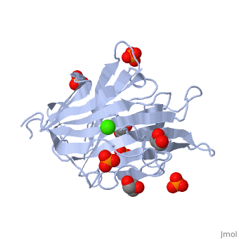
3atg
From Proteopedia
(Difference between revisions)
| Line 10: | Line 10: | ||
== Function == | == Function == | ||
[https://www.uniprot.org/uniprot/G1ED17_CELCE G1ED17_CELCE] | [https://www.uniprot.org/uniprot/G1ED17_CELCE G1ED17_CELCE] | ||
| - | <div style="background-color:#fffaf0;"> | ||
| - | == Publication Abstract from PubMed == | ||
| - | Endo-1,3-beta-glucanase from Cellulosimicrobium cellulans is composed of a catalytic domain and a carbohydrate-binding module. We have determined the X-ray crystal structure of the catalytic domain at a high resolution of 1.66A. The overall fold is a sandwich-like beta-jelly roll architecture like the enzymes in the glycoside hydrolase family 16. The substrate-binding cleft has a length and a width of ~28 and ~15A, respectively, which is thought to be capable of accommodating at least six glucopyranose units. Laminarihexaose was placed into the substrate-binding cleft, namely at the subsites +2 to -4 from the reducing end, and the complex structure was analyzed using molecular dynamics simulations (MD) and using a rotamer search of the pocket. During the MD simulations, the substrate fluctuated more than the enzyme, where the residues at the subsites toward the non-reducing end fluctuated more than those toward the reducing end. Little conformational change of the protein was observed for the subsites +1 and +2, indicating that the glucose's position could be tightly restricted inside the pocket. Substrate binding experiments using isothermal titration calorimetry showed that the binding affinity of laminaritriose was higher than that of laminaribiose and similar to those of other longer laminarioligosaccharides. Taken together, the substrates mainly bind to the subsites -1 to -3 with the highest affinity, while the part bound to the reducing end would be hydrolyzed. | ||
| - | |||
| - | Structural and thermodynamic characterization of endo-1,3-beta-glucanase: Insights into the substrate recognition mechanism.,Oda M, Inaba S, Kamiya N, Bekker GJ, Mikami B Biochim Biophys Acta. 2017 Dec 12;1866(3):415-425. doi:, 10.1016/j.bbapap.2017.12.004. PMID:29246508<ref>PMID:29246508</ref> | ||
| - | |||
| - | From MEDLINE®/PubMed®, a database of the U.S. National Library of Medicine.<br> | ||
| - | </div> | ||
| - | <div class="pdbe-citations 3atg" style="background-color:#fffaf0;"></div> | ||
==See Also== | ==See Also== | ||
*[[Glucanase|Glucanase]] | *[[Glucanase|Glucanase]] | ||
*[[Glucanase 3D structures|Glucanase 3D structures]] | *[[Glucanase 3D structures|Glucanase 3D structures]] | ||
| - | == References == | ||
| - | <references/> | ||
__TOC__ | __TOC__ | ||
</StructureSection> | </StructureSection> | ||
Current revision
endo-1,3-beta-glucanase from Cellulosimicrobium cellulans
| |||||||||||