User:Wayne Chang
From Proteopedia
(→Script Exercises) |
(→Script Exercises) |
||
| Line 15: | Line 15: | ||
<applet load='2qc8' size='300' frame='true' align='right' caption='Script Exercises' /> | <applet load='2qc8' size='300' frame='true' align='right' caption='Script Exercises' /> | ||
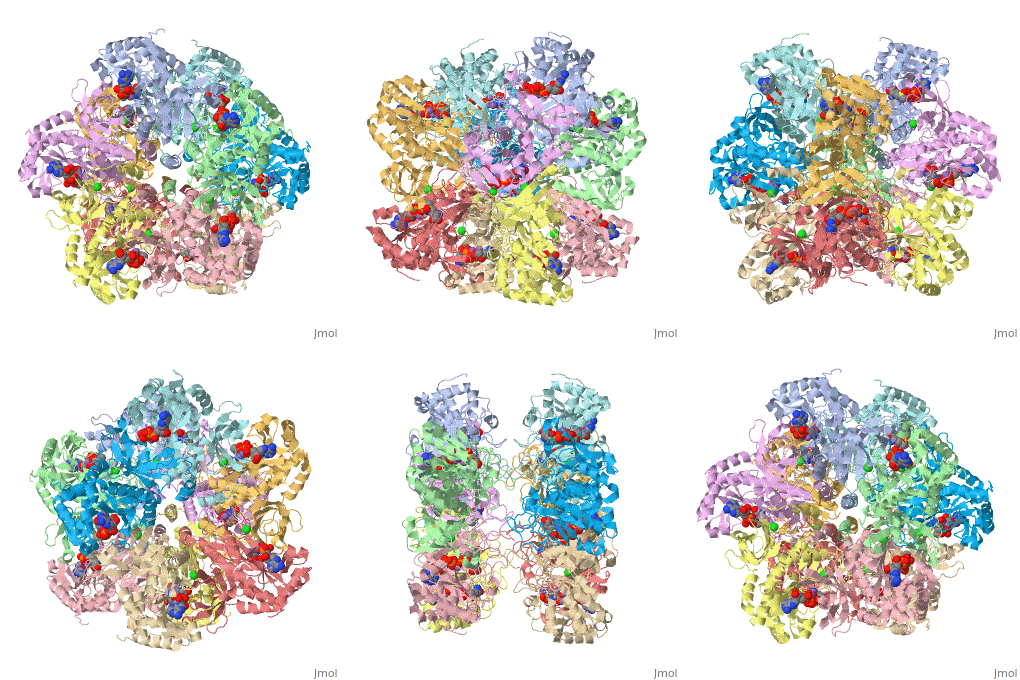
<scene name='User:Wayne_Chang/Glutamate_synthase/1'>Exercise 1: Backbone Trace with ligand</scene> | <scene name='User:Wayne_Chang/Glutamate_synthase/1'>Exercise 1: Backbone Trace with ligand</scene> | ||
| + | |||
Exercise shows a backbone trace of Glutamate Synthase which allows the ligands inside ADP, P3S, Cl- and Mn2+ to be seen. | Exercise shows a backbone trace of Glutamate Synthase which allows the ligands inside ADP, P3S, Cl- and Mn2+ to be seen. | ||
<scene name='User:Wayne_Chang/Glutamate_synthase/2'>Exercise 2: Ligand and Chain Selection with Labeling</scene> | <scene name='User:Wayne_Chang/Glutamate_synthase/2'>Exercise 2: Ligand and Chain Selection with Labeling</scene> | ||
| + | |||
Isolates chain A of Glutamate Synthase and labels the ligands for easy identification. | Isolates chain A of Glutamate Synthase and labels the ligands for easy identification. | ||
<scene name='User:Wayne_Chang/Glutamate_synthase/3'>Exercise 3: Active Site Residues</scene> | <scene name='User:Wayne_Chang/Glutamate_synthase/3'>Exercise 3: Active Site Residues</scene> | ||
| + | |||
Wire Structure of Active Site residues of chain A using information obtained from PDBsum entry for Glutamate Synthase. | Wire Structure of Active Site residues of chain A using information obtained from PDBsum entry for Glutamate Synthase. | ||
<scene name='User:Wayne_Chang/Glutamate_synthase/4'>Exercise 4: Going Solo</scene> | <scene name='User:Wayne_Chang/Glutamate_synthase/4'>Exercise 4: Going Solo</scene> | ||
| + | |||
Still picture of salt bridge between residue 63 of chain F and residue 319 of chain G. Bridge length is also provided in angstroms. | Still picture of salt bridge between residue 63 of chain F and residue 319 of chain G. Bridge length is also provided in angstroms. | ||
Revision as of 22:03, 15 November 2008
Contents |
Assignment 12: IVC: Ammonium Binding Site
Mapping the Ammonium binding site and explaining how it contributes to catalysis.
Chang, Wayne and Kaushal, Pankaj.
University of Maryland, Baltimore County (UMBC).
Script Exercises
|
Exercise shows a backbone trace of Glutamate Synthase which allows the ligands inside ADP, P3S, Cl- and Mn2+ to be seen.
Isolates chain A of Glutamate Synthase and labels the ligands for easy identification.
Wire Structure of Active Site residues of chain A using information obtained from PDBsum entry for Glutamate Synthase.
Still picture of salt bridge between residue 63 of chain F and residue 319 of chain G. Bridge length is also provided in angstroms.
Outline
-- Work in Progress --
Glutamine synthetase (GS) catalyzes the ATP dependent condensation of glutamate and ammonia, producing, glutamine, ADP, and an inorganic phosphate group.
Glutamate + ATP + NH3 → Glutamine + ADP + phosphate
Ammonium ion is thought to bind to GS at the monovalent cation binding site for Tl(+) and Cs(+) ions.
References
1. Liaw, S-H, et.al.,Discovery of the ammonium substrate site on glutamine synthetase, a third cation binding site Protein Sci. 1995 4: 2358-2365[1]

