This old version of Proteopedia is provided for student assignments while the new version is undergoing repairs. Content and edits done in this old version of Proteopedia after March 1, 2026 will eventually be lost when it is retired in about June of 2026.
Apply for new accounts at the new Proteopedia. Your logins will work in both the old and new versions.
Prion protein
From Proteopedia
| Line 2: | Line 2: | ||
=Structure of PrP<sup>C</sup>= | =Structure of PrP<sup>C</sup>= | ||
| - | PrP<sup>C</sup> has a natively unstructured N-terminal region, and a predominantly α-helical C-terminal region from residues ~120-230. | ||
| - | |||
| - | The N-terminal region can bind coper ions | ||
{{STRUCTURE_1hjm | PDB=1hjm | SCENE= }} | {{STRUCTURE_1hjm | PDB=1hjm | SCENE= }} | ||
| + | PrP<sup>C</sup> has a natively unstructured N-terminal region, and a predominantly α-helical C-terminal region from residues ~120-230, with a single disulfide bond. The presence of the N-terminus has little impact on the structure of the C-terminal domain <ref>1</ref>. | ||
The structure is highly conserved amongst mammals, and only differs slightly in birds, reptiles and amphibians. | The structure is highly conserved amongst mammals, and only differs slightly in birds, reptiles and amphibians. | ||
| - | + | ALthough having a similar overall fold, the X-ray structure of sheep PrP was dimeric | |
=Models of PrP<sup>Sc</sup> structure= | =Models of PrP<sup>Sc</sup> structure= | ||
| Line 17: | Line 15: | ||
=Genetic prion diseases= | =Genetic prion diseases= | ||
| - | A number of mutations in PrP have been identified which correlate with a high incidence of prion disease. To date, structural studies of all mutant PrP<sup>C</sup> have extremely similar structures to wild type PrP<sup>C</sup>, suggesting a kinetic basis for the difference in converting to PrP<sup>Sc</sup>. | + | A number of mutations in PrP have been identified which correlate with a high incidence of prion disease. The structure of HuPrP,E200K was determined nd shown to be To date, structural studies of all mutant PrP<sup>C</sup> have extremely similar structures to wild type PrP<sup>C</sup>, suggesting a kinetic basis for the difference in converting to PrP<sup>Sc</sup>. |
=Prion strains= | =Prion strains= | ||
| Line 30: | Line 28: | ||
* [[1QM2]] HuPrP residues 121-230 | * [[1QM2]] HuPrP residues 121-230 | ||
* [[1I4M]] HuPrP residues 119-226 (X-ray) | * [[1I4M]] HuPrP residues 119-226 (X-ray) | ||
| - | * [[1E1J]] HuPrP,M166V residues 125-228 | ||
| - | * [[1E1S]] HuPrP,S170N residues 125-228 | ||
| - | * [[1E1W]] HuPrP,R220K residues 125-228 | ||
* [[1FKC]] HuPrP,E200K residues 90-231 (genetic prion disease) | * [[1FKC]] HuPrP,E200K residues 90-231 (genetic prion disease) | ||
* [[1H0L]] HuPrP residues 121-230, with an additional disulphide bond analogous to the homolog Doppel | * [[1H0L]] HuPrP residues 121-230, with an additional disulphide bond analogous to the homolog Doppel | ||
| Line 46: | Line 41: | ||
=References= | =References= | ||
| + | {{Reflist}} <references/> | ||
| + | 1. | ||
Revision as of 11:44, 14 December 2008
The prion protein (PrP) is a cell surface glycoprotein. PrP can exist in two alternatively folded confirmations: the cellular isoform (PrPC) can undergo a structural conversion to a 'scrapie' or disease associated isoform termed PrPSc. Prion diseases such as Creutzfeldt Jakob disease (CJD) in people, and bovine spongiform encephalopathy (BSE) commonly known as "mad cow" disease, are characterterized by aggregates of PrPSc, which arise from autocatalytic refolding of PrPC in a template-dependent manner.
Contents |
Structure of PrPC
| |||||||||
| 1hjm, 1 NMR models () | |||||||||
|---|---|---|---|---|---|---|---|---|---|
| Related: | 1e1g, 1e1j, 1e1p, 1e1s, 1e1u, 1e1w, 1hjn | ||||||||
| |||||||||
| |||||||||
| Resources: | FirstGlance, OCA, RCSB, PDBsum | ||||||||
| Coordinates: | save as pdb, mmCIF, xml | ||||||||
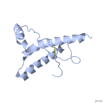

PrPC has a natively unstructured N-terminal region, and a predominantly α-helical C-terminal region from residues ~120-230, with a single disulfide bond. The presence of the N-terminus has little impact on the structure of the C-terminal domain [1].
The structure is highly conserved amongst mammals, and only differs slightly in birds, reptiles and amphibians.
ALthough having a similar overall fold, the X-ray structure of sheep PrP was dimeric
Models of PrPSc structure
Circular dichroism studies first demonstrated that PrPSc had very different proportions of α-helices and β-sheet to PrPC
There are a number of technical obstacles in determining the molecular structure of PrP(sup)Sc</sup>
Genetic prion diseases
A number of mutations in PrP have been identified which correlate with a high incidence of prion disease. The structure of HuPrP,E200K was determined nd shown to be To date, structural studies of all mutant PrPC have extremely similar structures to wild type PrPC, suggesting a kinetic basis for the difference in converting to PrPSc.
Prion strains
The strain phenomenon of prions ( ) was initially difficult to equate with the
Selected PrP structures
All structures determined by NMR unless otherwise specified
Human PrP
- 1QLX HuPrP residues 23-230
- 1QM0 HuPrP residues 90-230
- 1QM2 HuPrP residues 121-230
- 1I4M HuPrP residues 119-226 (X-ray)
- 1FKC HuPrP,E200K residues 90-231 (genetic prion disease)
- 1H0L HuPrP residues 121-230, with an additional disulphide bond analogous to the homolog Doppel
Other species PrPs
- XXXX Mouse PrP R
- 1B10 Syrian hamster PrP residues 90-231
- 1DWY Cow PrP residues 121-230
- 1UW3 Sheep PrP (X ray)
- XXXX Frog PrP
- XXXX Chicken PrP
- XXXX Turtle PrP
References
- ↑ 1
1.
Proteopedia Page Contributors and Editors (what is this?)
Kurt Giles, Joel L. Sussman, Jaime Prilusky, Michal Harel, Claudio Garutti, Eran Hodis
