This old version of Proteopedia is provided for student assignments while the new version is undergoing repairs. Content and edits done in this old version of Proteopedia after March 1, 2026 will eventually be lost when it is retired in about June of 2026.
Apply for new accounts at the new Proteopedia. Your logins will work in both the old and new versions.
User:Nathan Roy
From Proteopedia
| Line 43: | Line 43: | ||
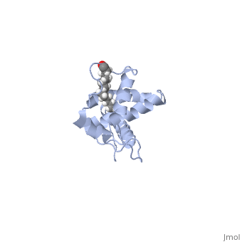
<applet load='1aum_dimer.pdb' size='300' frame='true' align='left' caption='FIGURE 3. Side-by-side structure of CA CTD' /> | <applet load='1aum_dimer.pdb' size='300' frame='true' align='left' caption='FIGURE 3. Side-by-side structure of CA CTD' /> | ||
| - | Once Gag is localized to discreet sites on the plasma membrane, multimerization of Gag takes place quite quickly , driven by the CA domain, and more specifically our focus here, the C-terminal domain of CA (CTD). There are four helices that contribute to the interaction of CA CTD with it's partner. A side-by-side interaction has been proposed (Figure 3), but many believe the forces involved in the side-by-side model are not great enough to account for the organization and structural stability of assembled Gag. Also, helix 1 of the CA CTD contains a very conserved region of residues within many retroviruses called the MHR (major homology region). In the side-by-side model, the MHR is not responsible for the dimer organization, therefore a model of the CA CTD dimer in which the MHR is responsible for organization of the CA CTD dimers has been sought. By making a single deletion of the Ala 177 residue (which lies in the loop between helix 1 and helix 2), the CA CTD domain adopts a domain-swapped conformation, in which the MHR of helix 1 is extended to contact helices 2,3, and 4 of the adjacent CA CTD domain (Figure 4). This domain swapping allows for a tighter binging of the CA CTD domains, and a stronger, and more rigid viral capsid. | + | Once Gag is localized to discreet sites on the plasma membrane, multimerization of Gag takes place quite quickly , driven by the CA domain, and more specifically our focus here, the C-terminal domain of CA (CTD). There are four helices that contribute to the interaction of CA CTD with it's partner. A side-by-side interaction has been proposed (Figure 3), but many believe the forces involved in the side-by-side model are not great enough to account for the organization and structural stability of assembled Gag. Also, helix 1 of the CA CTD contains a very conserved region of residues within many retroviruses called the MHR (major homology region)<scene name='User:Nathan_Roy/Mhr_side-by-side/1'>MHR region</scene>. In the side-by-side model, the MHR is not responsible for the dimer organization, therefore a model of the CA CTD dimer in which the MHR is responsible for organization of the CA CTD dimers has been sought. By making a single deletion of the Ala 177 residue (which lies in the loop between helix 1 and helix 2), the CA CTD domain adopts a domain-swapped conformation,<applet load='2ont_dimer.pdb' size='300' frame='true' align='right' caption='FIGURE 4. Domain swapped CA CTD' /> in which the MHR of helix 1 is extended to contact helices 2,3, and 4 of the adjacent CA CTD domain (Figure 4)(<scene name='User:Nathan_Roy/Mhr_domain_swapped/1'>Show MHR</scene>. This domain swapping allows for a tighter binging of the CA CTD domains, and a stronger, and more rigid viral capsid. |
Revision as of 21:59, 21 April 2009
User:Nathan Roy/Sandbox 1I am a graduate student at the University of Vermont, studying the dynamics of HIV-1 cell to cell transmission and HIV-1 induced syncytia formation. Our wonderful professor, Dr. Steven Everse, has commissioned us (his students from his BioChem 351 course) to create a page describing the structure of a protein that interests us. I have chosen the HIV-1 gag protein, and more specifically, the MA and CA domains.
HIV-1 Gag
The HIV-1 Gag protein is the major structural protein required for virus assembly. It is synthesized as a polyprotein in the cytosol of an infected cell, and contains four functional segments; MA, CA (NTD and CTD), NC, and p6. The NC region is flanked by two "spacer" segments, denoted SP1 and SP2. The polyprotein is all alpha helical, except the NC region, which is composed of two RNA interacting zinc knuckle domains. Gag is often referred to as an "assembly machine", because expression of Gag alone is sufficient to produce budding virus-like particles (VLP's), due to multimerization of roughly 2000 Gag molecules per virion. Here, we will take a closer look at the MA, CA, and NC domains, and how the structural components of these domains aid in the assembly of virus particles. Viral particles can be classified as immature (pre-budding and non-infectious), and mature (post-budding and infectious), and this exchange is mediated by the HIV-1 protease(LINK). Upon viral budding, Gag is cleaved by the HIV-1 protease at multiple sites, thus possibly changing many of the structural interactions that make up the "immature" particle. For simplicity, we will only be discussing the immature formation of Gag on the plasma membrane of infected cells, as it coordinates organized viral budding.
Matrix (MA)
|
|
Capsid (CA)
|
|
Nucleocapsid (NC)
|
