File list
From Proteopedia
| Name | User | Size | Description | |
|---|---|---|---|---|
| 12:45, 16 March 2010 | MPSceneVersion01.spt (file) | Eric Martz | 2 KB | |
| 13:14, 15 March 2010 | Beta_m110_130.pdb (file) | Tilman Schirmer | 3 KB | (beta strand with all phi,psi = -110,130) |
| 02:46, 12 March 2010 | Table.JPG (file) | Bhagiradhi Somalanka | 125 KB | |
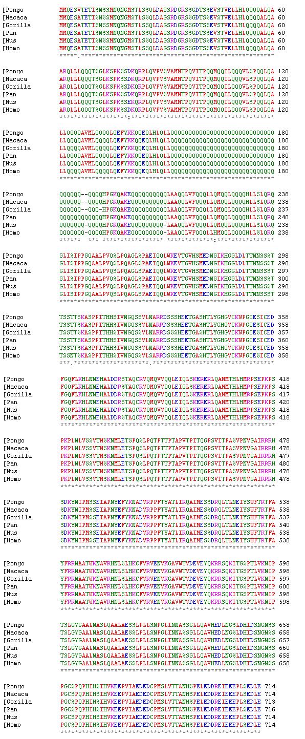
| 02:17, 12 March 2010 | Alignment2.JPG (file) | Liz Thomas | 346 KB | (Alignment of foxp2 for human, chimp, gorilla, rhesus monkey, and mouse. ) |
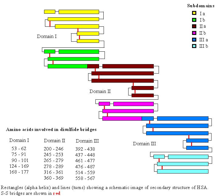
| 18:01, 11 March 2010 | HSA-disulfide_bridges_drawing.JPG (file) | Irene Becerra | 61 KB | (This is a representation of the disulfide bridges, alpha helix, and domains/subdomains of HSA.) |
| 12:04, 11 March 2010 | Kyc.gif (file) | Alexander Berchansky | 168 KB | (Morph) |
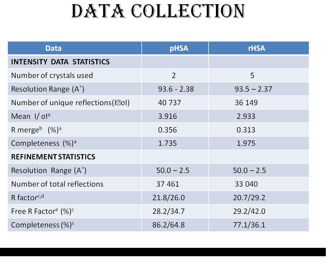
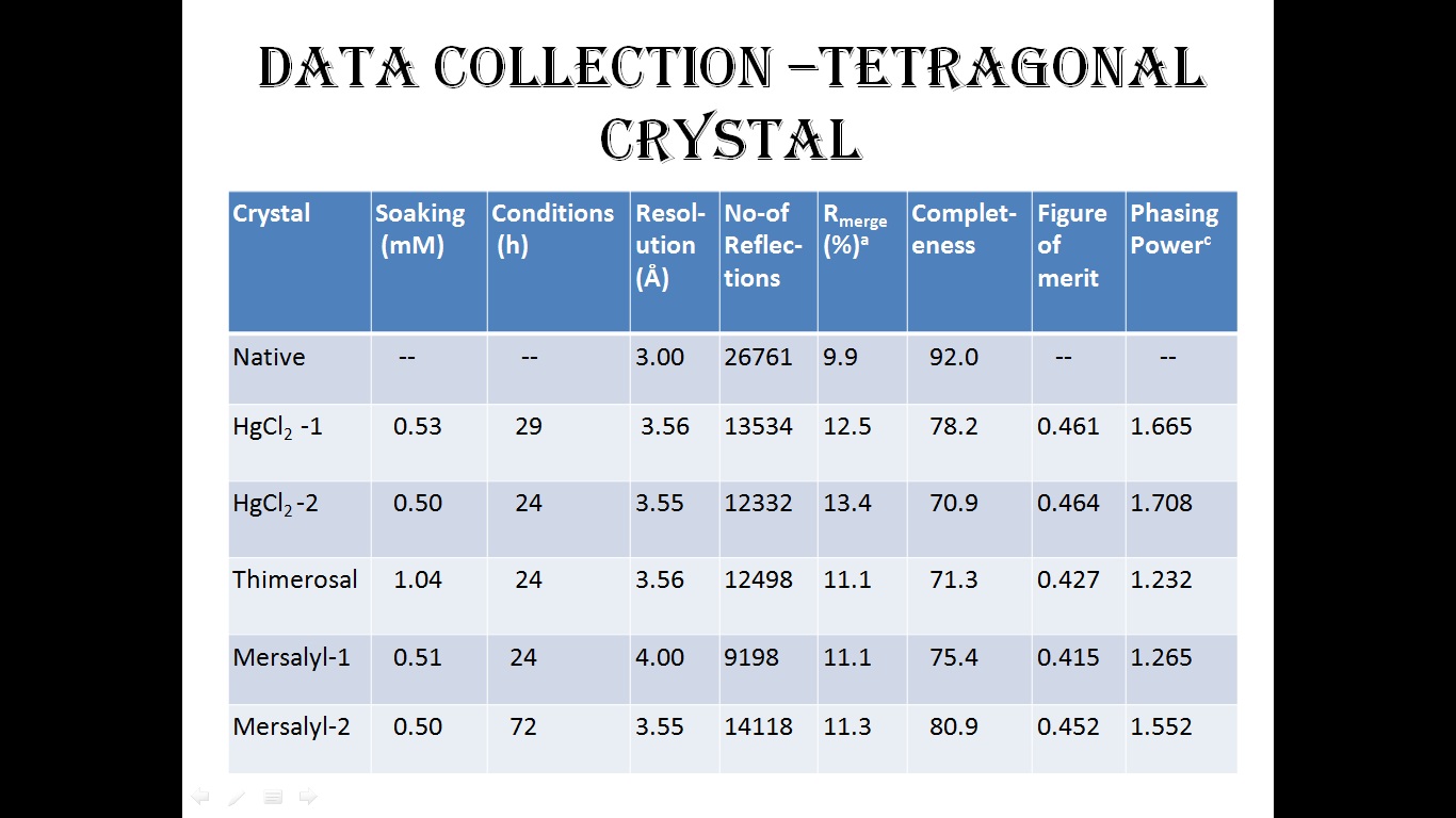
| 18:25, 10 March 2010 | Data_collection.jpg (file) | Neeharika Pothuri | 164 KB | |
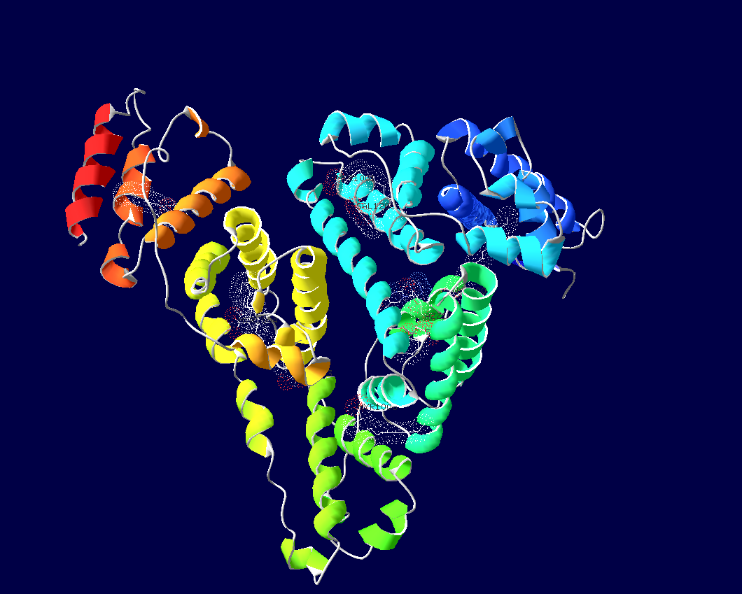
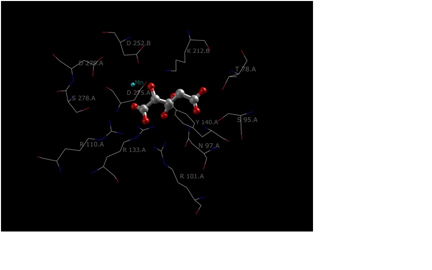
| 04:59, 10 March 2010 | 3b9m_3D_image_with_ligands.png (file) | Bhagiradhi Somalanka | 204 KB | |
| 23:42, 9 March 2010 | Hab3.pdb (file) | Yuan-Ping Pang | 580 KB | (Model 3 of HAB•BoNTAe) |
| 23:42, 9 March 2010 | Hab2.pdb (file) | Yuan-Ping Pang | 576 KB | (Model 2 of HAB•BoNTAe) |
| 23:14, 9 March 2010 | Hab1.pdb (file) | Yuan-Ping Pang | 573 KB | (Model 1 of HAB•BoNTAe) |
| 19:07, 9 March 2010 | 1N4K.pdb (file) | Shannon King | 239 KB | |
| 04:07, 9 March 2010 | StapPep.pdb (file) | Craig T Martin | 9 KB | |
| 04:05, 9 March 2010 | Peptide12mer_R_PentS_Oct_11RCM.pdb (file) | Craig T Martin | 9 KB | (Stapled peptide) |
| 14:21, 8 March 2010 | 1ea5_2010.pdb (file) | Eran Hodis | 465 KB | |
| 22:24, 7 March 2010 | AdhD_with_three_cofactors.pdb (file) | Tommie Hata | 192 KB | (AdhD) |
| 22:19, 7 March 2010 | AdhD_with_three_cofactors_no_H.pdb (file) | Tommie Hata | 192 KB | (AdhD) |
| 05:04, 7 March 2010 | Table1.jpg (file) | Bhagiradhi Somalanka | 165 KB | |
| 04:52, 7 March 2010 | Table.gif (file) | Bhagiradhi Somalanka | 91 KB | |
| 21:13, 6 March 2010 | Ligand.jpg (file) | Neeharika Pothuri | 256 KB | |
| 20:39, 6 March 2010 | With_ligand.jpg (file) | Neeharika Pothuri | 256 KB | |
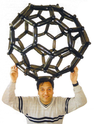
| 15:07, 6 March 2010 | Balloon_buckyball.png (file) | Eric Martz | 190 KB | (From http://balloonmolecules.com/) |
| 19:54, 5 March 2010 | Dragon_genetics_concord.org.png (file) | Eric Martz | 8 KB | (Snapshot from Dragon Genetics, from Concord.Org, specifically [http://geniquest.portal.concord.org/preview/]) |
| 22:45, 4 March 2010 | Gramicidin_in_bilayer.pdb.gz (file) | Eric Martz | 66 KB | (See header of this PDB file for provenance: Crouzy et al, Biophys J 67:1370, 1994. Gramicidin theoretical model in hydrated bilayer of phosphatidyl ethanolamine.) |
| 15:06, 3 March 2010 | 3iio.jpg (file) | OCA | 45 KB | (Source PDB http://www.rscb.org/pdb) |
| 12:11, 3 March 2010 | 3kyd.png (file) | OCA | 423 KB | (Source Jena Library http://www.fli-leibniz.de/IMAGE.html) |
| 09:17, 3 March 2010 | 2nd.png (file) | Jasper Small | 28 KB | |
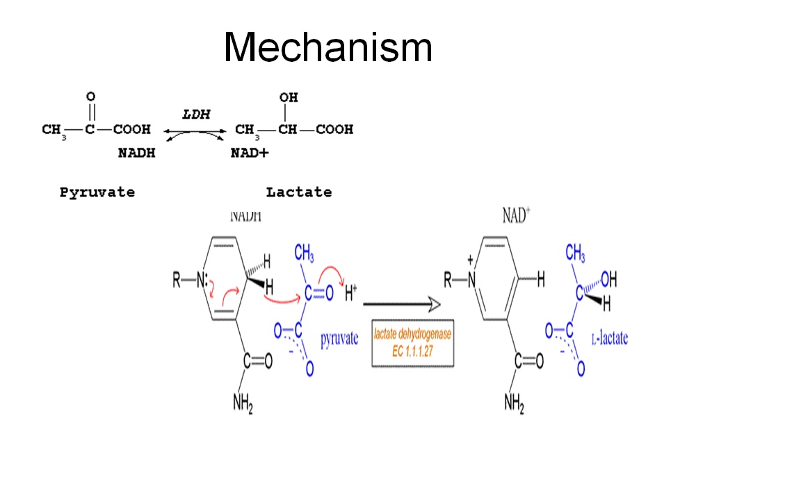
| 03:03, 3 March 2010 | LDH_reaction.jpeg (file) | Jasper Small | 65 KB | (Rxn shown) |
| 16:54, 2 March 2010 | Expand_residues.jvxl (file) | Karl Oberholser | 15 KB | (Selected AC1 site, Pro-cap, Leu-lock, Recoil before computing surface.) |
| 13:45, 2 March 2010 | 4pfk.mmol (file) | Zach Westrick | 667 KB | |
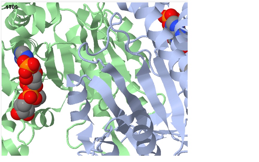
| 20:21, 1 March 2010 | Human_isocitrate.jpg (file) | Michael Nobbe | 111 KB | |
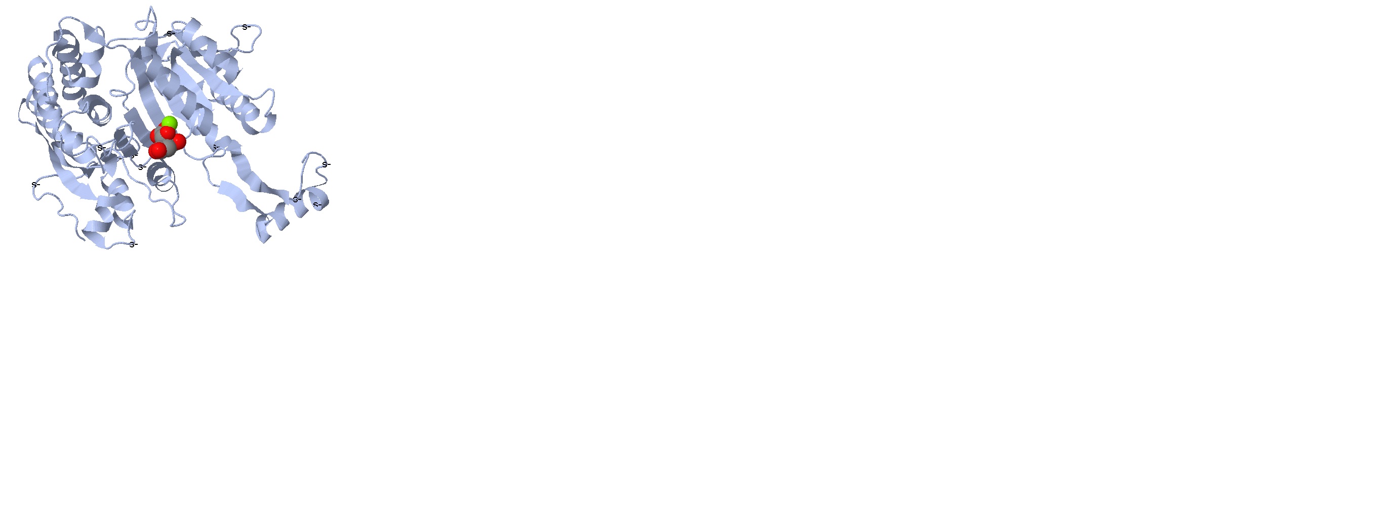
| 20:20, 1 March 2010 | E_coli_phosphorylation_active_site_isocitrate.jpg (file) | Michael Nobbe | 134 KB | |
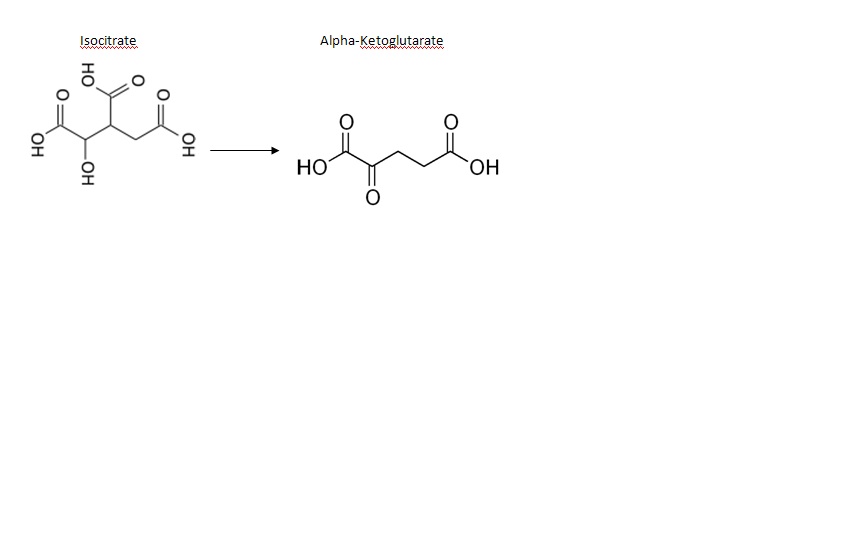
| 20:20, 1 March 2010 | Chemicals_isocitrate.jpg (file) | Michael Nobbe | 21 KB | |
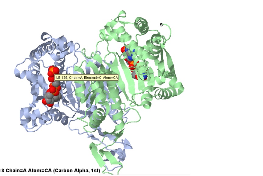
| 20:20, 1 March 2010 | Active_site_isocitrate.jpg (file) | Michael Nobbe | 136 KB | |
| 20:09, 1 March 2010 | A.jpg (file) | Michael Nobbe | 35 KB | |
| 18:25, 1 March 2010 | 1nek.mmol (file) | Michael Vick | 2 KB | |
| 17:47, 1 March 2010 | Aconitase.JPG (file) | Anthony Noles | 13 KB | |
| 17:02, 1 March 2010 | QuinoneMechanism.gif (file) | Michael Vick | 11 KB | (Image 3: Reduction of ubiquinone to ubiquinol (from Wikimedia Commons)) |
| 16:57, 1 March 2010 | S.D.Oxidation_of_Succinate_E1cb.gif (file) | Michael Vick | 10 KB | (Image 2: Oxidation of succinate to fumarate via E1cb elimination (from Wikimedia Commons)) |
| 16:53, 1 March 2010 | S.D.Oxidation_of_Succinate_E2.gif (file) | Michael Vick | 7 KB | (Image 1: Oxidation of succinate to fumarate through E2 elimination) |
| 03:14, 1 March 2010 | Active_site_movement.jpg (file) | Bogdan Stancu | 222 KB | (Substrate induced movement) |
| 23:39, 28 February 2010 | 2ald.mmol (file) | Austin Drake | 766 KB | |
| 23:32, 28 February 2010 | Align.jpg (file) | Bogdan Stancu | 79 KB | (Phosphoglucoisomerase multiple alignment) |
| 22:53, 28 February 2010 | 1iat.mmol (file) | Bogdan Stancu | 679 KB | (Phosphoglucoisomerase dimer) |
| 19:47, 28 February 2010 | 1one.mmol (file) | Cory Tiedeman | 515 KB | |
| 03:29, 28 February 2010 | 2cts.mmol (file) | Daniel Eddelman | 474 KB | |
| 03:15, 28 February 2010 | 1cts.mmol (file) | Daniel Eddelman | 455 KB | |
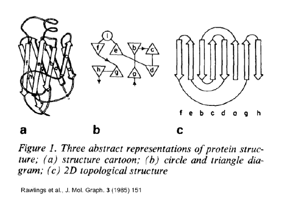
| 10:15, 25 February 2010 | Prealbumin_topology2.png (file) | Tilman Schirmer | 66 KB | |
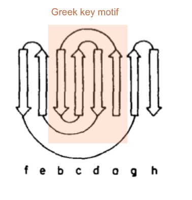
| 10:10, 25 February 2010 | Prealbumin_topology_greek_key1.png (file) | Tilman Schirmer | 40 KB | (from Rawlings et al., J. Mol. Graph. 3 (1985) 151) |
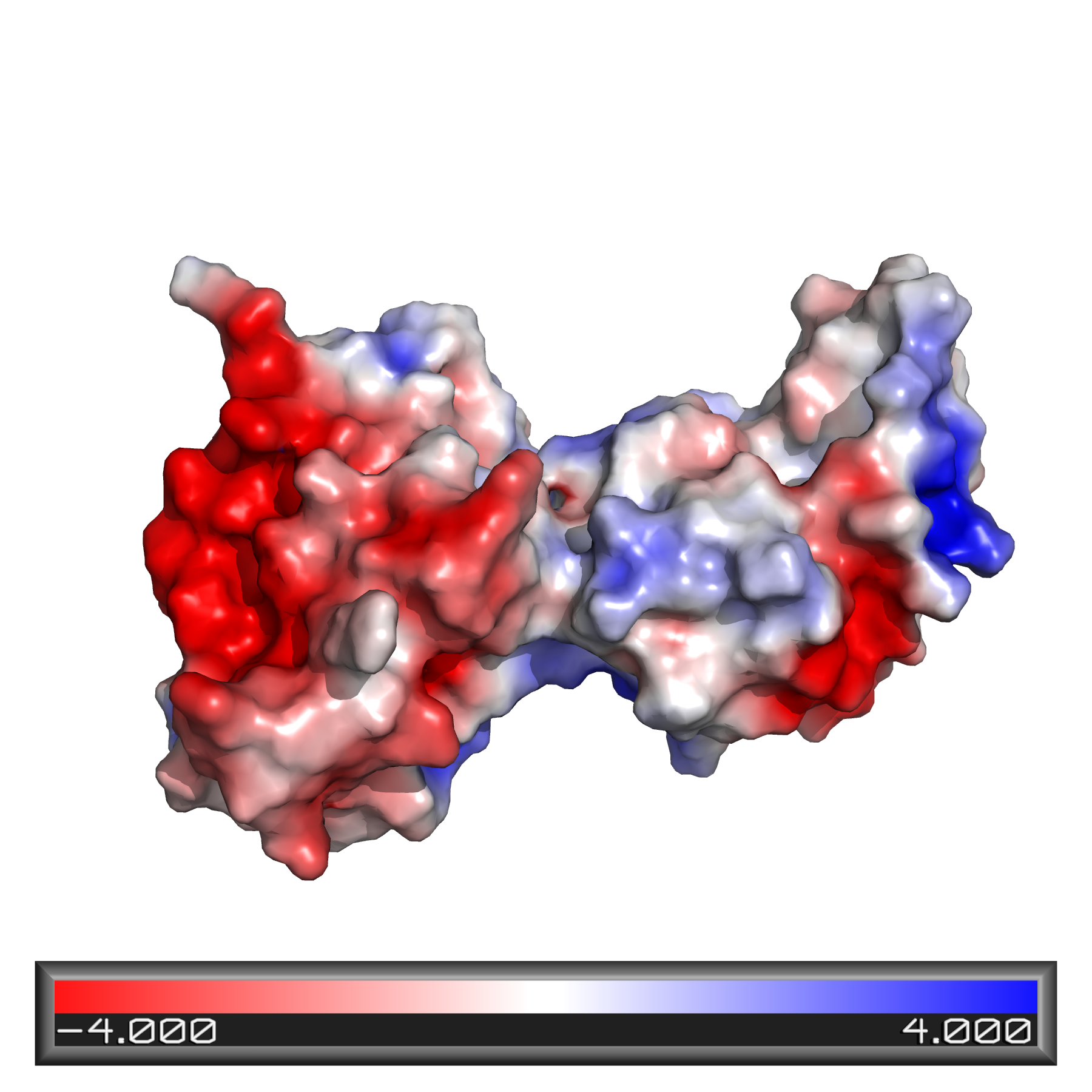
| 08:58, 21 February 2010 | 1KT0_Domain12_Elc.png (file) | Orly Dym | 871 KB |
First page |
Previous page |
Next page |
Last page |
