This old version of Proteopedia is provided for student assignments while the new version is undergoing repairs. Content and edits done in this old version of Proteopedia after March 1, 2026 will eventually be lost when it is retired in about June of 2026.
Apply for new accounts at the new Proteopedia. Your logins will work in both the old and new versions.
2emt
From Proteopedia
| |||||||
| , resolution 2.80Å | |||||||
|---|---|---|---|---|---|---|---|
| Related: | 1J19, 1GC6, 1GC7, 2D11, 2D10, 2D2Q, 2EMS
| ||||||
| Resources: | FirstGlance, OCA, PDBsum, RCSB | ||||||
| Coordinates: | save as pdb, mmCIF, xml | ||||||
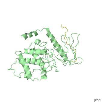
Crystal Structure Analysis of the radixin FERM domain complexed with adhesion molecule PSGL-1
Overview
P-selectin glycoprotein ligand-1 (PSGL-1), an adhesion molecule with O-glycosylated extracellular sialomucins, is involved in leukocyte inflammatory responses. On activation, ezrin-radixin-moesin (ERM) proteins mediate the redistribution of PSGL-1 on polarized cell surfaces to facilitate binding to target molecules. ERM proteins recognize a short binding motif, Motif-1, conserved in cytoplasmic tails of adhesion molecules, whereas PSGL-1 lacks Motif-1 residues important for binding to ERM proteins. The crystal structure of the complex between the radixin FERM domain and a PSGL-1 juxtamembrane peptide reveals that the peptide binds the groove of FERM subdomain C by forming a beta-strand associated with strand beta5C, followed by a loop flipped out towards the solvent. The Motif-1 3(10) helix present in the FERM-ICAM-2 complex is absent in PSGL-1 given the absence of a critical Motif-1 alanine residue, and PSGL-1 reduces its contact area with subdomain C. Non-conserved positions are occupied by large residues Met9 and His8, which stabilize peptide conformation and enhance groove binding. Non-conserved residues play an important role in compensating for loss of binding energy resulting from the absence of conserved residues important for binding.
About this Structure
2EMT is a Protein complex structure of sequences from Mus musculus. Full crystallographic information is available from OCA.
Reference
Structural basis of PSGL-1 binding to ERM proteins., Takai Y, Kitano K, Terawaki S, Maesaki R, Hakoshima T, Genes Cells. 2007 Dec;12(12):1329-38. PMID:18076570
Page seeded by OCA on Mon Mar 31 02:51:13 2008
