The RNase H2 ribonuclease complex is a heterotrimeric endoribonuclease responsible for the major ribonuclease H activity in mammalian cells. In mouse, the complex is encoded by 3 genes located on chromosomes 8 (Rnaseh2a), 14 (Rnaseh2b) and 19 (Rnaseh2c)[1]. This enzyme specifically cleaves the 3’O-Phosphate bond of RNA in a DNA/RNA hybrids to produce 5’ phosphate and 3’hydroxyl ends.
Biological role
Ribonucleases H are the only known enzymes, able to degrade the RNA strand of a DNA/RNA hybrid in a sequence-nonspecific way.
There are two types of RNase H (RNases H1 and RNases H2) classified according to their sequence conservation and substrate preference. Currently, three types of RNA/DNA hybrids are known: simple RNA/DNA duplexes (Figure 1A), RNA•DNA/DNA hybrids (Figure 1B), and DNA•RNAfew•DNA/DNA hybrids (Figure 1C). RNases H2 is totally able to cleave a single ribonucleotide embedded in a double strand DNA (DNA• RNAfew •DNA/DNA type) when RNases H1 require at least 4 ribonucleotides. This ability and their high expression in proliferating cells suggest that RNases H2 are involved in DNA repair and replication[2].
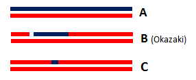
Figure 1 : Three types of RNA/DNA hybrids
Indeed, ribonucleotides are wrongly incorporated into DNA during DNA replication at a frequency of about 2 ribonucleotides per kb. With such frequency, these errors are by far the most abundant threat of DNA damaging. Hence, a correction is essential to the preservation of DNA integrity: the most common correction mechanism involves RNases H2 and is called Ribonucleotide Excision Repair (RER). The incorporation of ribonucleotides in DNA produce DNA•RNAfew•DNA/DNA hybrids from which the few misincorporated ribonucleotides can be removed by an RNase H2[3].
This repair activity is guided by the interaction between C-terminus of RNase H2B protein and the DNA clamp PCNA. This interaction occurs through a hydrophobic conserved peptide motif called the PCNA interaction peptide PIP (PIP-box: Residues 294 to 301 MKSIDTFF of H2B protein) that interacts with a hydrophobic groove near the PCNA C-terminus. This interaction allows RNase H2 to scan DNA for misincorporated ribonucleotides which makes the Ribonucleotide Excision Repair more efficient[4].
Furthermore, in vitro studies have shown that RNases H2 is likely to be involved in the removal of RNA primer from Okazaki fragment produced during the synthesis of the lagging strand in DNA replication since Okazaki fragment are RNA•DNA/DNA hybrids (Figure 1B)[4].
RNases H2 activity is crucial in mammalian cells, for instance a mutation in human RNase H2 causes Aicardi-Goutières syndrome. This syndrome is an auto-inflammatory disorder that may be the consequence of an increased production of incorrect nucleic acid by-products during DNA replication[4].
Structure
A heteromeric complex
It has been shown that the Mammalian RNase complex is a heteromeric complex formed by 3 distinct proteins: , and . H2A protein is the catalytic subunit and H2B/H2C proteins are auxiliary subunits: they are structural domains that facilitate cohesion of the complex[5].
The first domain structure of the complex, H2A, contains 301 amino acids, almost as H2B protein which computes 308 amino acids. H2C protein is the smallest subunit: it only has 166 amino acids.
Each of these proteins adopts various secondary structures with β-strands and α-helices:
  H2A protein has 12 α-helices, 11 β-strands and 3 turns[6],
  H2B molecule computes 8 α-helices, 7 β-strands and 3 turns[7],
  H2C subunit consists of 5 α-helices, 8 β-strands and 2 turns[8].
Several interactions between the subunits
H2C protein is found in the middle of the elongated complex structure, flanked by H2A and H2B proteins on the ends.
The complex is stabilized by the intimately interwoven architecture of H2B and H2C: The N-terminal region of H2B protein (amino acids 1-92) weaves together with H2C domain to form 3 β-barrels, called [9]. This triple barrel is formed from a total of and produces a pseudo-2-fold axis of symmetry along the central barrel. Also, it permits to leave the mostly α-helical C-terminal region of H2B available for potential interactions with other protein (for example the PCNA protein). Finally, it has been found that the motif provides a platform for securely binding the H2A protein: the side and end of the first barrel in the subcomplex H2B/H2C form a with amino acids 197-258 in the C-terminal region of H2A protein. This interface is mainly composed of hydrophobic residues
Interactions with nucleic acids
It has been proved that the position of RNA/DNA complex in the active site cleft is determined by several favorable electrostatic interactions between the nucleic acid and positively charged amino acids of the protein[2]

