Sandbox Home

From Proteopedia

Revision as of 16:15, 30 September 2025 by Joel L. Sussman (Talk | contribs)
Jump to: navigation, search
       ISSN 2310-6301
     
       As life is more than 2D, Proteopedia helps to bridge the gap between 3D structure & function of biomacromolecules
     
     
       Proteopedia presents this information in a user-friendly way as a collaborative & free 3D-encyclopedia of proteins & other biomolecules.
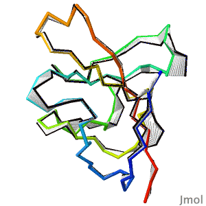
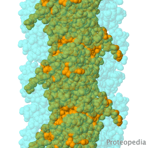
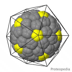
     
Selected Research Pages In Journals Education
About this image
BREAKTHROUGH in protein structure prediction!

by Eric Martz
After decades of slow progress by many groups, in 2020, AlphaFold2 proved able to accurately predict the detailed structures of two-thirds of single protein domains from their amino acid sequences. Pictured is AlphaFold2's prediction for the ORF8 protein of SARS-CoV-2 (black), compared with a subsequently published X-ray crystallographic structure (colors). ORF8 contributes to virulence in COVID-19.
>>> Visit this page >>>

About this image
Geobacter pili: surprising function.

Y Gu, V Srikanth, AI Salazar-Morales, R Jain, JP O'Brien, SM Yi, RK Soni, FA Samatey, SE Yalcin, NS Malvankar. Nature 2021 doi: 10.1038/s41586-021-03857-w
Geobacter pili were long thought to be electrically conductive protein nanowires composed of PilA-N. Nanowires are crucial to the energy metabolism of bacteria flourishing in oxygen-deprived environments. To everyone's surprise, in 2019, the long-studied nanowires were found to be linear polymers of multi-heme cytochromes, not pili. The first cryo-EM structure of pili (2021) reveals a filament made of dimers of PilA-N and PilA-C, shown. Electrical conductivity of pili is much lower than that of cytochrome nanowires. Evidence suggests that PilA-NC filaments are periplasmic pseudopili crucial for exporting cytochrome nanowires onto the cell surface, rather than the pili serving as nanowires themselves.

>>> Visit I3DC Interactive Visualizations >>>

About this image
Virus Capsid Geometry

The Capsid of a virus is its outer shell or "skin". Viruses have evolved intricate and elegant ways to assemble capsid protein chains into complete, usually spherical capsids, often with icosahedral symmetry. Pictured is an extremely simplified model of a capsid, where a single enlarged atom represents each of the 360 protein chains in the capsid of the Simian Virus 40 (SV40), a member of a group of cancer-causing viruses that has been extensively researched for decades.

>>> See more animations and explanation >>>

About Contact Hot News Table of Contents Structure Index Help
Personal tools