Main Page

From Proteopedia

Revision as of 11:17, 21 October 2018 by Joel L. Sussman (Talk | contribs)
Jump to: navigation, search
Often it is difficult to utilize the wealth of information found in 3D biomacromolecular structures. Proteopedia's goal is to present structure/function information on these molecules in a user-friendly manner to a broad scientific audience. </td></tr>
ISSN 2310-6301

As life is more than 2D, Proteopedia helps to bridge the gap between 3D structure & function of biomacromolecules

Selected Pages Art on Science Journals Education
About this image
Bacteria float with nano-balloons.

ST Huber, D Terwiel, WH Evers, D Maresca, AJ Jakobi. Preprint 2022 doi: 10.1101/2022.05.08.489936
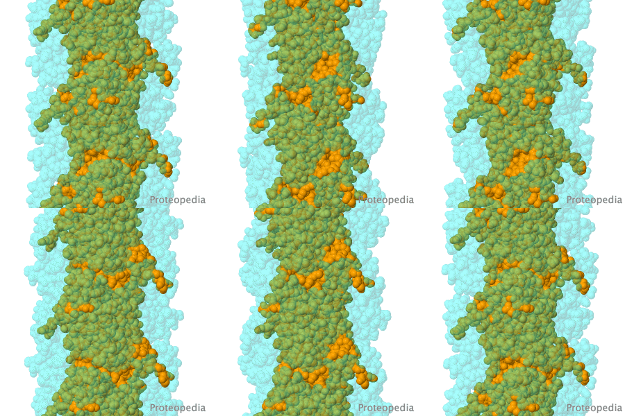
Many kinds of bacteria and archaea control their buoyancy to move to optimal positions in liquid environments. They do this by making nano-compartments called "gas vesicles", long "pipes" with closed ends filled with gases. In 2022, gas vesicle structure was solved, revealing self-assembling thin-walled cylinders of remarkable strength with gas-permeable pores and water-repelling (hydrophobic) interiors. Building on this structural knowledge, gas vesicles are being engineered to serve as biosensors that report via ultrasound.

>>> Visit I3DC Interactive Visualizations >>>

About this image
Opening a Gate to Human Health

by Alice Clark (PDBe)
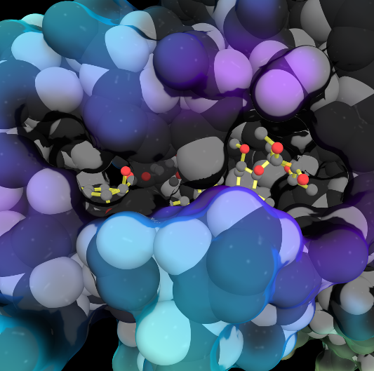
In the 1970s, an exciting discovery of a family of medicines was made by the Japanese scientist Satoshi Ōmura. One of these molecules, ivermectin, is shown in this artwork bound in the ligand binding pocket of the Farnesoid X receptor, a protein which helps regulate cholesterol in humans. This structure showed that ivermectin induced transcriptional activity of FXR and could be used to regulate metabolism.

>>> Visit this page >>>

About this image
Structural flexibility of the periplasmic protein, FlgA, regulates flagellar P-ring assembly in Salmonella enterica.

H Matsunami, YH Yoon, VA Meshcheryakov, K Namba, FA Samatey. Scientific Reports 2016 doi: 10.1038/srep27399
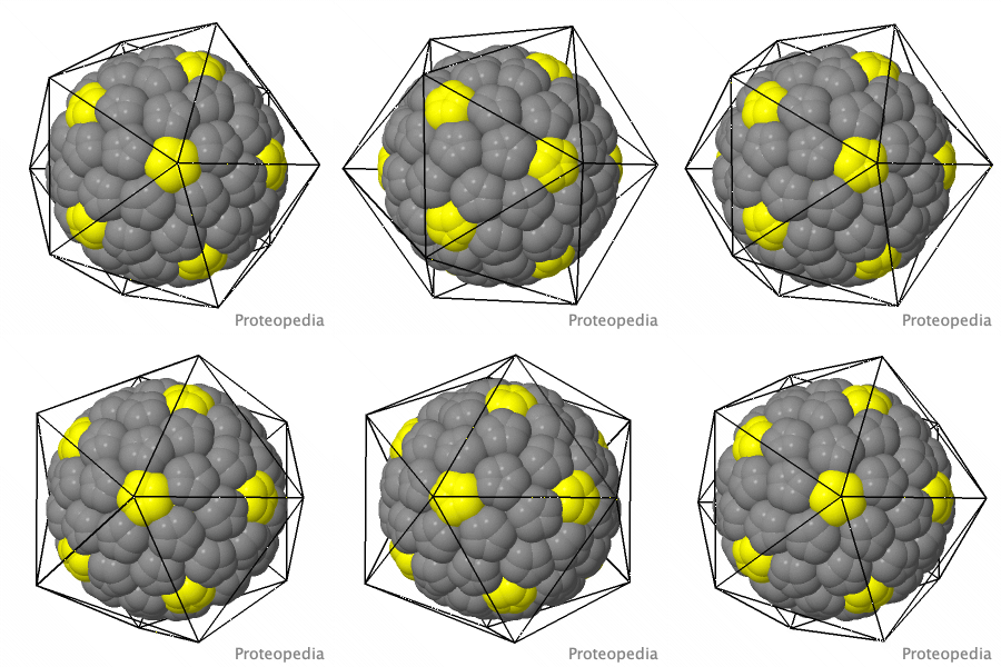
A periplasmic flagellar chaperone protein, FlgA, is required for P-ring assembly in bacterial flagella of taxa such as Salmonella enterica or Escherichia coli. Here we present the open and closed crystal structures of FlgA from Salmonella enterica serovar Typhimurium, grown under different crystallization conditions. An intramolecular disulfide cross-linked form of FlgA caused a dominant negative effect on motility of the wild-type strain.

>>> Visit this I3DC complement >>>

About this image
You Are What You Eat!

Above is an integral membrane protein that takes up, into your intestinal cells, orally consumed peptide nutrients and drugs. Its lumen-face (top) opens and binds peptide or drug (small solid object in the center), then closes, while its cytoplasmic face (bottom) opens to release its cargo into the intestinal cell, which passes it on to the blood circulation.

>>> See more animations and explanation >>>

How to add content to Proteopedia

Video Guides

Who knows ...

List of Art on Science pages

About Interactive 3D Complements - I3DCs

List of I3DCs

How to get an I3DC for your paper

Teaching strategies using Proteopedia

Examples of pages for teaching

How to add content to Proteopedia

About Contact Table of Contents Structure Index Help

Proteopedia Page Contributors and Editors (what is this?)

Joel L. Sussman, Jaime Prilusky

Personal tools