Deubiquitinating enzymes or Deubiquitinases (DUBs) are enzymes with an ubiquitin-dependent action. More than a hundred DUBs genes exist in humans, making it a very diverse protein and allowing targeted action. Their main role is to cleave ubiquitin bound to a substrate, often a protein. Ubiquitin, bound to the substrate, allows it to regulate its degradation by the proteasome or lysozyme, influences its cellular localisation or modulates the activity with another protein. [1][2]
Generalities
Function
Deubiquitinases are key enzymes belonging to the vast group of proteases, allowing the degradation of ubiquitin of proteins. These enzymes are thus implicated in the regulation of protein degradation. Indeed, when a protein is going to be degraded, an enzymatic cascade will add a poly-ubiquitin fragment to the protein. This mechanism is called ubiquitination(fr) (en). Following this step, mono or poly-ubiquitin is removed from the protein which has been degraded, by deubiquitinase. [3]
Families
Deubiquitinases belong to the protease family. This family is divided into five classes, according to the nature of the amino acid composition of their active site carrying out the catalysis: serine protease, cysteine proteases, acid proteases, metalloproteases, threonine proteases. DUBs belong to only two of these families: metalloproteases and cysteine proteases.
Among the cysteine proteins, four subfamilies can be described according to their catalytic domains: ubiquitin-specific proteases (USP), les Ubiquitin C-terminal hydrolases (UCH), Otubain proteases (OTU) and Machado-joseph disease proteases (MJD). The deubiquitinases belonging to the family of metalloproteases all have a JAMM catalytic domain (JAB1/MPN/Mov34 metalloenzyme).
Within these two families, DUBs are classified into subfamilies according to the differences in their amino acid sequences surrounding the catalytically active amino acid residues. [4]
Localization
The localization depends on the DUB we consider. However, the majority of DUBs are found in the nucleus, plasma membrane or/and in secretory and endocytic pathways. For example, in the ubiquitin-specific proteases family, the USP21 is mostly associated with microtubules and the centrosome. Thus this deubiquitinase depends on all the physiological mechanisms involving the microtubules. [5]
Structure
The overall structure
3TMP is the catalytic domain of human deubiquitinase DUBA in complex with ubiquitin aldehyde. It is a 8 chain structure with sequence from Human. Indeed, 3TMP is made of these two macromolecules : OTU domain-containing protein 5 (also named DUBA or OTUD5) and Polyubiquitin-C, which is the ubiquitin aldehyde. It is also made of two small molecules which are the phosphoserine (SEP) and the amino-acetaldehyde (GLZ), they are L-peptide links. [6]
Impact of phosphorylation on DUB activity
Evidence shows that phosphorylation influences activity of the enzyme. Phosphorylated serine seems to have the most influence on the activity of the enzyme . The phosphorylation of this nucleotide is crucial and the protein won't work if it's not.
In fact, this part bends to welcome the protein to be deubiquitinased.
Catalytic domain
It is the catalytic domain which defines the family of the DUB. Indeed, DUBs belonging to the family of cysteine proteases have a catalytic site composed of two or three amino acids (dyads or triads). When the catalytic site is active, it may contain cysteine, histidine, aspartate or asparagine residues. In the case of metalloproteases, the active site is composed of a zinc ion and amino acids such as histidine, aspartate and serine. [7]
The studied structure shows both
Residues present in the catalytic site of DUBs are often in a non-functional orientation when the substrate is absent. Thus, when the substrate binds to the catalytic site of the enzyme, the site undergoes rearrangement and takes on a functional conformation. [8] The substrate opens and closes to allow the entry of the protein to be deubiquitinased.
The enzyme take this configuration thanks to This is why phosphorylation is so important to the function of the enzyme. The phosphate group forms many links between substrate ubiquitin and a segment of the OTU domain. This is rare among the known structures of deubiquitinases. Phosphorylation-driven conformational change ressembles the one of kinases.
Biological role
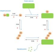
The role of DUBs is in the ubiquitin pathway. The modifications made by DUBs are post-translational modifications. Thus, DUBs have different functions related to ubiquitin:
A : maturation of ubiquitin. When ubiquitin molecules are synthesized, they are not in free form. Thus, DUBs are essential for the generation of free monomers from precursors. The degradation of precursors is carried out by several DUBs belonging to the USPs class.
B : cleavage between protein and mono-ubiquitin and regulation of the poly-ubiquitin chain. DUBs also have a regulatory activity because they allow the elimination of ubiquitin chains mistakenly conjugated to substrates.
C : cleavage between protein and poly-ubiquitin chain. Once the protein has been degraded by the proteasome or autophagolysosome, DUBs allow the polyubiquitin chain to be separated from the protein.
D : recycling of ubiquitin. Certain deubiquitinases allow the separation of polyubiquitin chains in order to release ubiquitin monomers. [9]

Figure 1 : Role of DUBs in the ubiquitin pathways
Disease
The involvement of deubiquitinases in diseases is still poorly understood. However, it is known that they play a role in various physiological processes, particularly in the case of cancers. [10]
In fact, DUBs have a role in the mechanism involved in histone modification and so have influence on tumor development and progression. For instance, in gastric cancer, DUBs are regulated upwards and DUBs are related to tumor size. [11]
Otubain 1
The enzyme Otubain 1 is a deubiquitinase belonging to the Otubain family of proteases. The role of OTUB1 is not yet clearly defined, some studies show the correlation between tumour growth and OTUB1 while others show no involvement or suppression of the tumour by this enzyme. This suggests that the effects of OTUB1 depend on the stage and the tumour itself. However, the therapeutic targeting of OTUB1 could be used for patients with various tumours, since the enzyme is found in many tissues and has a high level of cell expression. [12]

