Image:Domains of beta catenin.jpg
From Proteopedia

No higher resolution available.
Domains_of_beta_catenin.jpg (324 × 236 pixel, file size: 26 KB, MIME type: image/jpeg)
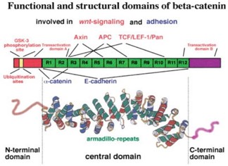
Fig. 1 – Structure of β-catenin with highlighted binding sites for interacting partner: top – domain structure of β-catenin, bottom – tertiary structure of β-catenin. Red inscription – interacting partners of β-catenin participating in Wnt signal pathway, blue inscription – interacting partners of β-catenin participating in forming of adherent junctions, R1-R12 – armadillo repetitions, each armadillo repetition is represented by red, green and blue α-helix in bottom part of the picture [1]
- ↑ Schneider SQ, Finnerty JR, Martindale MQ. Protein evolution: structure-function relationships of the oncogene beta-catenin in the evolution of multicellular animals. J Exp Zool B Mol Dev Evol. 2003 Feb 15;295(1):25-44. doi: 10.1002/jez.b.6. PMID:12548541 doi:http://dx.doi.org/10.1002/jez.b.6
File history
Click on a date/time to view the file as it appeared at that time.
| Date/Time | User | Dimensions | File size | Comment | |
|---|---|---|---|---|---|
| (current) | 21:04, 27 April 2022 | Kristína Galvánková (Talk | contribs) | 324×236 | 26 KB | Fig. 1 – Structure of β-catenin with highlighted binding sites for interacting partner: top – domain structure of β-catenin, bottom – tertiary structure of β-catenin. Red inscription – interacting partners of β-catenin participating in Wnt sig |
| 17:17, 26 April 2022 | Kristína Galvánková (Talk | contribs) | 324×236 | 26 KB | Fig. 1 – Structure of β-catenin with highlighted binding sites for interacting partner: top – domain structure of β-catenin, bottom – tertiary structure of β-catenin. Red inscription – interacting partners of β-catenin participating in Wnt sig |
- Edit this file using an external application
See the setup instructions for more information.
Links
The following pages link to this file:
