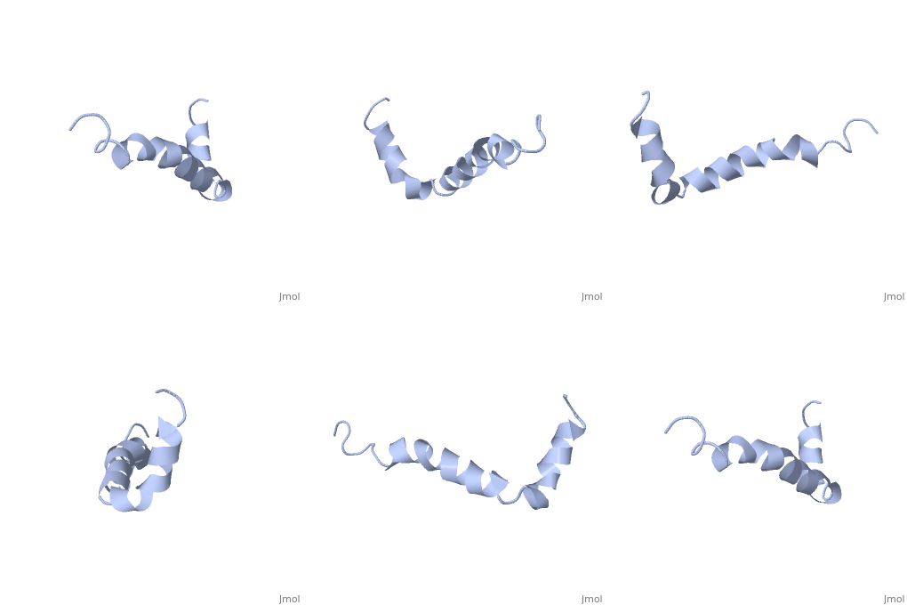
Amyloid beta
From Proteopedia
| |||||||||||
Neurotoxicity
Generation of Radicals
Amyloid beta generates reactive oxygen species and induces oxidative stress and inflammation. When complexed with Cu(II) amyloid beta becomes a moderate oxidant that is capable of creating hydrogen peroxide in the presence of air and a reducing agent (such as ascorbate).[3]
Toxicity in the Vasculature
A build up of amyloid beta results in the oxidative stress, caspase activation, mitochondrial dysfunction and DNA damage. Once amyloid beta produces superoxide it can react with nitrogen oxide and cause vasoconstriction, decreasing the blood supply to the brain.[3]
Pore Formation
Amyloid beta can adhere to endothelial cell walls and create lesions, eventually a large deposit will cause cerebral hemorrhage. The pores cause loss of calcium homeostasis and an influx of Ca2+ into neurons.[3]
Interaction with tau
Tau proteins function to stabilize microtubules [[2]]. Association between Tau proteins and amyloid beta results in dissociation of tau from microtubules which collapses the axonal structure leading to the death of neurons.[3]
Binding ApoE
Apolipoprotein E is normally involved in lipoprotein metabolism and transfer but it has been shown to play a role in the development of Alzheimer's. ApoE4 has the most affinity for amyloid beta and uses the low-density lipoprotein-related protein receptor to internalize amyloid beta into neurons. It also promotes the production of amyloid beta by stimulating APP recycling. It is also theorized that apolipoproteins promote the aggregation of amyloid beta into toxic oligomers.[3]
Binding ABAD
Amyloid beta-peptide binding alcohol dehydrogenase (ABAD) is an enzyme belonging to a family of short chain dehyndrogenase/reductases.[5] When amyloid beta binds to ABAD in the mitochondria there is increased mitochondrial dysfunction, increased reactive oxygen species and oxidative stress.[3] It has been found that by inhibiting amyloid beta from binding ADAB, using a decoy peptide, oxygen consumption is increased and the toxic effect are decreased. [6]
Binding Catalase
Catalase normally functions to convert hydrogen peroxide to hydrogen and water. If amyloid beta complexes with catalase at residues 31-35 it can no longer breakdown the toxic molecule. Is it thought that the inactivation of catalase is due to the insertion of the sulfur side chain of methionine into the catalytic site.[3]
Prevention and Treatment
Only preliminary studies have begun on the prevention of Alzheimer's. No true prevention has been determined but potential forms of prevention and risk factors have been identified. One promising preventative measure has been anti-inflammatory drugs. By taking anti-inflammatory drugs, it is thought that patients with arthritis may decrease/prevent brain inflammation which may prevent some of the stress that leads to Alzheimer's disease.[7] Major risk factors seem to center around diet and exercise. Both mental and physical exercise are likely to play a role in maintaining mental stability. For instance, it has been shown that individuals in midlife with raised systolic blood pressure and high serum cholesterol concentration, and in particular the combination of these risks, increase the risk of Alzheimer's disease in later life[8].
Two major approaches have been taken to treating Alzheimer's; inhibiting the formation of APP and reducing the neurotoxic effects of amyloid beta itself. The most promising treatment is the prevention of the enzymes responsible for creating APP, AF267B. This is a muscarinic receptor that activates alpha-secretase and reduces tau pathology.[3] Very recently it was discovered the loss of active JNK associated with the absence of both MKK4 and MKK7 protects neurons against amyloid beta-induced toxicity and JNK signaling is required for amyloid plaque formation in vivo.[9] Some potential targets for treatment include inhibiting beta sheet formation, creating molecules with high affinity for the self recognition region to prevent oligomerization, and tau pathology.
References
- ↑ 1.0 1.1 1.2 1.3 Crescenzi O, Tomaselli S, Guerrini R, Salvadori S, D'Ursi AM, Temussi PA, Picone D. Solution structure of the Alzheimer amyloid beta-peptide (1-42) in an apolar microenvironment. Similarity with a virus fusion domain. Eur J Biochem. 2002 Nov;269(22):5642-8. PMID:12423364
- ↑ Vlassenko AG, Benzinger TL, Morris JC. PET amyloid-beta imaging in preclinical Alzheimer's disease. Biochim Biophys Acta. 2011 Nov 12. PMID:22108203 doi:10.1016/j.bbadis.2011.11.005
- ↑ 3.00 3.01 3.02 3.03 3.04 3.05 3.06 3.07 3.08 3.09 3.10 3.11 3.12 Rauk A. Why is the amyloid beta peptide of Alzheimer's disease neurotoxic? Dalton Trans. 2008 Mar 14;(10):1273-82. Epub 2008 Feb 12. PMID:18305836 doi:10.1039/b718601k
- ↑ Funke SA. Detection of Soluble Amyloid-beta Oligomers and Insoluble High-Molecular-Weight Particles in CSF: Development of Methods with Potential for Diagnosis and Therapy Monitoring of Alzheimer's Disease. Int J Alzheimers Dis. 2011;2011:151645. Epub 2011 Nov 2. PMID:22114742 doi:10.4061/2011/151645
- ↑ Amyloid β-Peptide-binding Alcohol Dehydrogenase Is a Component of the Cellular Response to Nutritional Stress: Yan, Shi Du et al. (2000). DOI: 10.1074/jbc.M000055200
- ↑ Inhibition of Amyloid-beta (A beta) peptide-binding alcohol dehydrogenase-A beta interaction reduces A beta accumulation and improves mitochondrial function in a mouse model of Alzheimer's disease: Yao, Jen et al. (2006). [1]
- ↑ McGeer PL, Schulzer M, McGeer EG. Arthritis and anti-inflammatory agents as possible protective factors for Alzheimer's disease: a review of 17 epidemiologic studies. Neurology. 1996 Aug;47(2):425-32. PMID:8757015
- ↑ Kivipelto M, Helkala EL, Laakso MP, Hanninen T, Hallikainen M, Alhainen K, Soininen H, Tuomilehto J, Nissinen A. Midlife vascular risk factors and Alzheimer's disease in later life: longitudinal, population based study. BMJ. 2001 Jun 16;322(7300):1447-51. PMID:11408299
- ↑ Attenuating GABAA Receptor Signaling in Dopamine Neurons Selectively Enhances Reward Learning and Alters Risk Preference in Mice: Parker, Jones G et al. (2011). DOI: 10.1523/JNEUROSCI.1715-11.2011

