Human Cardiac Troponin C
From Proteopedia
| |||||||||
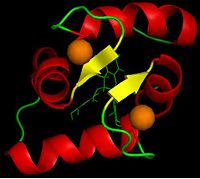
| NMR structure of human troponin C showing Ca, complex with green tea polyphenol, 2kdh | |||||||||
|---|---|---|---|---|---|---|---|---|---|
| Ligands: | , | ||||||||
| Gene: | TNNC1, TNNC (Homo sapiens) | ||||||||
| |||||||||
| |||||||||
| Resources: | FirstGlance, OCA, RCSB, PDBsum | ||||||||
| Coordinates: | save as pdb, mmCIF, xml | ||||||||
2KDH is the Solution Structure of Human Cardiac Troponin C in complex with the Green Tea Polyphenol : (-)-epigallocatechin-3-gallate. It is a 1 chain structure of sequence from Homo sapiens. That is the PDB name for the solution structure of Human Cardiac Troponin C in complex with the green tea Polyphenol (-)-epigallocatechin-3-gallate. Troponin C is a protein which can bind with calcium. That’s a subunit from a 3 subunited protein which is called Troponin. This protein is found in muscles. For example, heart muscle contraction is regulated by Ca2+ binding of troponin C to the thin filament.[1] In cardiovascular diseases, the myofilament response to Ca2+ is often altered. Compounds that rectify this perturbation are of considerable interest as therapeutics. Plant flavonoids have been found to provide protection against a variety of human illnesses such as cancers, infections, and heart diseases. (-)-Epigallocatechin gallate (EGCg), the prevalent flavonoid in green tea is one of those plant flavonoids.
Contents |
From the binding of Ca2+ with TnC to the muscular contraction
The troponin C is a part of the troponin complex which is composed of three proteins: troponin C, troponin I and troponin T. The troponin C is involved in the process of muscular contraction. There are some residues which interact with two Ca2+ (one Ca2+ binds with AC1 domain of the molecule). That generates a change of conformation in the troponin complex. In fact, the troponin C is bound with the two others troponin (TnI and TnT) and a change of conformation in the troponin C (induces by the Ca2+ binding) generates a change of conformation of the troponin complex. So the troponin I releases the site on the actin which can now bind the myosin. The myosin binds the actin site and has a ATPase activity which permits finally the muscular contraction.
This molecule is very polar,in fact there are lot of aspartic acid and glutamic acid which provide negative charges and several lysin and arginin which confer positive charges. That molecule can so interract a lot with its environment.
Structure of troponin C and interactions with the EGCg
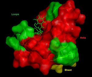
The troponin C is a 72 residues protein organized with and which are anti-parallel. This structure appears on the Ramachandran plot, where can be observed a high concentration of residues (amino acids) in the energy level located in: Psi 0-(-45), Phi -90-(-45), which is the level of energy the most favorable to the constitution of helices. On the contrary, only few residues are in the level of energy corresponding to the sheet:Psi 90-180, Phi -135-(-45), actually there are only 2 little sheets. There are some residues in: Psi 0-90, Phi 45-90, which are involved in loops permitting bounds between sheets and helices. Four of those residues are glycins which permit those loops because of their low steric dimensions. In fact, glycin with it radical chain composed of one hydrogen allows easily change of direction of the protein. It's one of amino acids the most privileged in those kinds of structure.
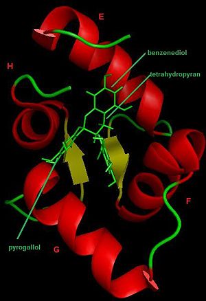
There are which form an hydrophobic pocket.EGCg interacts with AC3 hydrophobic domain of the molecule and binds with AC2 domain. But there are wich have no interactions with that molecule and this suggets that the binding of EGCg is near of the hydrophobic pocket rather than deep within the pocket and it induces a small structural "opening". The opening degree of TnC is described by the inter-helical angles between helices E and F and between helices G and H.
As said just before, EGCg makes contacts exclusively to hydrophobic residues that line the surface of TnC. Actually it binds near the surface of helix E, so near the N-terminus of TnC, with tetrahydropyran and benzenediol. The pyrogallol ring stays near the C-terminus of TnC, which explains the large chemical shift perturbations of some residues of the .
Moreover, EGCg can bind with the TnC-2Ca2+ or TnC-2Ca2+-TnI and forms a ternary complex, which increases the binding potential.
Therapy application
Common treatment schemes of heart failure modify levels of cytosolic Ca2+. This provides immediate improvement in heart function, but it can lead to serious side effects if used for an extended period of time. Drugs that alter the Ca2+ sensitivity of the thin filament rather than the cytosolic Ca2+ concentration, provide a safer alternative. An increase in Ca2+ sensitivity would be beneficial for the treatment of heart failure, whereas the use of Ca2+ desentizers may provide protection against the development of hypertrophic cardiomyopathy (HCM).
EGCg has a role as Ca2+ desensitizer and is particularly interesting in regards to treatment for HCM because that is a scavenger of radicals and this may help treat or prevent HCM by sequestering reactive oxygen species as well as by inhibiting ATPases activity (this prevents binding of Ca2+ so there is no ATPase activity).
Green tea contains that EGCg polyphenol and it can then act as a modulator of heart contraction through its interaction with TnC. So the use of green tea can help in some heart diseases, and can be an alternative for a start for a treatment at home .
3D structures of troponin C
References
- ↑ Robertson IM, Li MX, Sykes BD. Solution structure of human cardiac troponin C in complex with the green tea polyphenol, (-)-epigallocatechin 3-gallate. J Biol Chem. 2009 Aug 21;284(34):23012-23. Epub 2009 Jun 20. PMID:19542563 doi:10.1074/jbc.M109.021352
The journal of biological chemistry
Proteopedia Page Contributors and Editors (what is this?)
Alicia Daeden, Céline Challemel, Audrey Chabrat, Michal Harel


