This old version of Proteopedia is provided for student assignments while the new version is undergoing repairs. Content and edits done in this old version of Proteopedia after March 1, 2026 will eventually be lost when it is retired in about June of 2026.
Apply for new accounts at the new Proteopedia. Your logins will work in both the old and new versions.
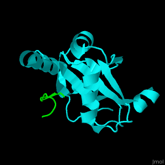
Microtubule-associated protein
From Proteopedia
| |||||||||||
References
- ↑ Maccioni RB, Cambiazo V. Role of microtubule-associated proteins in the control of microtubule assembly. Physiol Rev. 1995 Oct;75(4):835-64. PMID:7480164
- ↑ Halpain S, Dehmelt L. The MAP1 family of microtubule-associated proteins. Genome Biol. 2006;7(6):224. PMID:16938900
- ↑ Lebouvier T, Scales TM, Williamson R, Noble W, Duyckaerts C, Hanger DP, Reynolds CH, Anderton BH, Derkinderen P. The microtubule-associated protein tau is also phosphorylated on tyrosine. J Alzheimers Dis. 2009;18(1):1-9. doi: 10.3233/JAD-2009-1116. PMID:19542604 doi:http://dx.doi.org/10.3233/JAD-2009-1116
- ↑ Buligescu L, Lenkei R, Ciontea M, Dan EM. [Significance of anti-albumin antibodies in chronic liver disease]. Rev Med Interna Neurol Psihiatr Neurochir Dermatovenerol Med Interna. 1977, Jul-Aug;29(4):363-70. PMID:22919
- ↑ Turtola LO. Enamel microhardness and fluoride uptake underneath fermenting and non-fermenting artificial plaque. Scand J Dent Res. 1977 Sep;85(6):373-9. PMID:22924
- ↑ Schraen-Maschke S, Dhaenens CM, Delacourte A, Sablonniere B. Microtubule-associated protein tau gene: a risk factor in human neurodegenerative diseases. Neurobiol Dis. 2004 Apr;15(3):449-60. PMID:15056452 doi:http://dx.doi.org/10.1016/j.nbd.2003.12.009
- ↑ Ichimura Y, Kumanomidou T, Sou YS, Mizushima T, Ezaki J, Ueno T, Kominami E, Yamane T, Tanaka K, Komatsu M. Structural basis for sorting mechanism of p62 in selective autophagy. J Biol Chem. 2008 Aug 15;283(33):22847-57. Epub 2008 Jun 4. PMID:18524774 doi:http://dx.doi.org/M802182200
Proteopedia Page Contributors and Editors (what is this?)
Michal Harel, Alexander Berchansky, Joel L. Sussman, Jaime Prilusky
