Sandbox Reserved 598
From Proteopedia
| Line 15: | Line 15: | ||
== Analyzing and Discovering Jak2 Structure == | == Analyzing and Discovering Jak2 Structure == | ||
| - | In determining the three-dimensional structure of the Jak2 protein, three primary methods of analysis were used; protein expression and purification, crystallization, and x-ray data collection. <ref> Lucet, I., Fantino, E., & Styles, M. (2005). The structural basis of janus kinase 2 inhibition by a potent and specific pan-janus kinase inhibitor. Blood, 107, 176-183. doi: 10.1182/blood-2005-06-2413 http://bloodjournal.hematologylibrary.org/content/107/1/176.full.pdf </ref> | + | In determining the three-dimensional structure of the Jak2 protein, three primary methods of analysis were used; protein expression and purification, crystallization, and x-ray data collection. <ref> Lucet, I., Fantino, E., & Styles, M. (2005). The structural basis of janus kinase 2 inhibition by a potent and specific pan-janus kinase inhibitor. Blood, 107, 176-183. doi: 10.1182/blood-2005-06-2413 http://bloodjournal.hematologylibrary.org/content/107/1/176.full.pdf </ref> In the protein expression and purification the Jak2 residue was cloned using pFastBac which uses “two promoters in a single vector for expression of two proteins simultaneously in insect cells”. <ref> http://www.invitrogen.com/1/1/14896-pfastbac-dual.html </ref> The bacmid DNA with the kinase insert was then isolated and put into cells of insect army worms. The cells were then grown, lysed and centrifuged after which the protein was then incubated, separated with gel filtration and fractions were taken for crystallization trials. In these crystallization trials, the protein residue was used to grow crystals via hanging drop vapor-diffusion. The purified protein complex was then mixed with solutions which subsequently formed crystals after one to three days. The crystalized protein was then flash frozen and the structure determined via molecular replacement and the AmoRe program. <ref> ) Lucet, I., Fantino, E., & Styles, M. (2005). The structural basis of janus kinase 2 inhibition by a potent and specific pan-janus kinase inhibitor. Blood, 107, 176-183. doi: 10.1182/blood-2005-06-2413 http://bloodjournal.hematologylibrary.org/content/107/1/176.full.pdf </ref> |
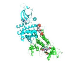
| - | + | <Structure load='2B7A' size='300' frame='true' align='right' caption='Janus Kinase 2' scene='3-D Structure of Jak2, as seen on Protein Data Bank' /> | |
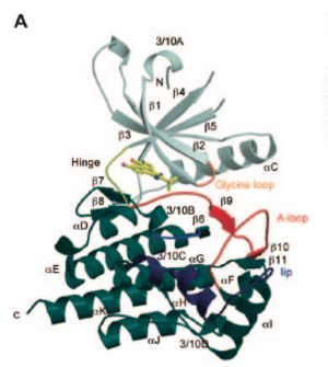
| - | + | [[Image:Jak2structure.png|thumb|300px|left|Structure of Jak2 as determined by Dr. Isabelle S. Lucet et al]] | |
| - | <Structure load='2B7A' size='300' frame='true' align='right' caption='Janus Kinase 2' scene=' | + | The structure of Jak 2 can be broken down into seven separate components, as seen in the pictured diagram to the left. |
Revision as of 18:54, 14 April 2013
| This Sandbox is Reserved from Feb 1, 2013, through May 10, 2013 for use in the course "Biochemistry" taught by Irma Santoro at the Reinhardt University. This reservation includes Sandbox Reserved 591 through Sandbox Reserved 599. |
To get started:
More help: Help:Editing |
Janus Kinase 2 (Jak2)
Background
Janus Kinase 2 is a non-receptor janus kinase, a protein which is part of the tyrosine kinases. These group of kinases are the primary intracellular mediators of cytokine signaling and are involved in the control of cellular growth. As a non-receptor kinase, Jak 2 has a cytoplasmic enzyme which catalyzes the transfer of a phosphate group through phosphorylation to the tyrosine residue in the protein. Such an enzyme plays a crucial role in regulating various cellular functions by switching on or off additional enzymes within the cell. [1] Such phosphorylation is a reversible process, and used in many different pathways as a method to control cellular activity. However kinases like Jak2, have enzymes which add phosphate groups to hydroxyl side chains as can be seen in the diagram. [2] 
Jak2 was given its name "Janus" after the two-faced Roman God "Janus" who was known as the custodian of the universe and the God of new beginnings. [3] The abbreviation 'Jak' is commonly referred to as 'just another kinase' as, when it was first discovered, the kinase's role was not yet fully understood. [4]
Analyzing and Discovering Jak2 Structure
In determining the three-dimensional structure of the Jak2 protein, three primary methods of analysis were used; protein expression and purification, crystallization, and x-ray data collection. [5] In the protein expression and purification the Jak2 residue was cloned using pFastBac which uses “two promoters in a single vector for expression of two proteins simultaneously in insect cells”. [6] The bacmid DNA with the kinase insert was then isolated and put into cells of insect army worms. The cells were then grown, lysed and centrifuged after which the protein was then incubated, separated with gel filtration and fractions were taken for crystallization trials. In these crystallization trials, the protein residue was used to grow crystals via hanging drop vapor-diffusion. The purified protein complex was then mixed with solutions which subsequently formed crystals after one to three days. The crystalized protein was then flash frozen and the structure determined via molecular replacement and the AmoRe program. [7]
|
The structure of Jak 2 can be broken down into seven separate components, as seen in the pictured diagram to the left.


