SUMO
From Proteopedia
| Line 11: | Line 11: | ||
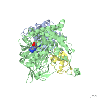
The <scene name='3kyc/Al/2'>structural alignment</scene> of the crystal structures for human SUMO E1 in complex with SUMO adenylate (AMSN) and tetrahedral intermediate (AVSN) analogues revealed opened conformation (<font color='orange'><b>SUMO1 in orange</b></font>, <font color='blue'><b>SAE1 colored in blue</b></font>, and <font color='darkviolet'><b>other domains in darkviolet</b></font>) and closed conformation (<font color='yellow'><b>SUMO1 in yellow</b></font>, <font color='cyan'><b>SAE1 colored in cyan</b></font>, and <font color='magenta'><b>other domains in magenta</b></font>), respectively. In the <scene name='3kyc/Al/7'>open conformation</scene> ([[3kyc]]) the distance between Cys domain (including Cys173) and mimic of the acyl adenylate intermediate AMSN is very long, while in the <scene name='3kyc/Al/6'>closed conformation</scene> ([[3kyd]]), the catalytic Cys173 is posioned near AVSN and SUMO1, so the overall structure revealed dramatic rearrangement. This large conformational change forms the <scene name='3kyc/Al/8'>E1~SUMO1-AVSN tetrahedral intermediate analogue</scene>. | The <scene name='3kyc/Al/2'>structural alignment</scene> of the crystal structures for human SUMO E1 in complex with SUMO adenylate (AMSN) and tetrahedral intermediate (AVSN) analogues revealed opened conformation (<font color='orange'><b>SUMO1 in orange</b></font>, <font color='blue'><b>SAE1 colored in blue</b></font>, and <font color='darkviolet'><b>other domains in darkviolet</b></font>) and closed conformation (<font color='yellow'><b>SUMO1 in yellow</b></font>, <font color='cyan'><b>SAE1 colored in cyan</b></font>, and <font color='magenta'><b>other domains in magenta</b></font>), respectively. In the <scene name='3kyc/Al/7'>open conformation</scene> ([[3kyc]]) the distance between Cys domain (including Cys173) and mimic of the acyl adenylate intermediate AMSN is very long, while in the <scene name='3kyc/Al/6'>closed conformation</scene> ([[3kyd]]), the catalytic Cys173 is posioned near AVSN and SUMO1, so the overall structure revealed dramatic rearrangement. This large conformational change forms the <scene name='3kyc/Al/8'>E1~SUMO1-AVSN tetrahedral intermediate analogue</scene>. | ||
</StructureSection> | </StructureSection> | ||
| - | __NOTOC__ | ||
{{Clear}} | {{Clear}} | ||
Revision as of 10:51, 20 August 2014
| |||||||||||

For better understanding of the difference between these two conformations you can see this morph (generated by using POLYVIEW-3D: http://polyview.cchmc.org/polyview3d.html; reload/refresh this page to restart this movie). Of note, in contrast to the previous figure, the same domains of these two structures (3kyc and 3kyd) are colored in the same colors (SUMO1 in yellow, SAE1 colored in blue and other domains in darkviolet). The catalytic Cys173 is shown in the spacefill representation and colored green, AMSN (or AVSN) are shown in the spacefill representation and colored in CPK colors.
Contents |
3D Structures of SUMO
Updated on 20-August-2014
SUMO
2k8h – SUMO – NMR – Trypanosoma brucei
1u4a – hSUMO-3 (mutant) – NMR – human
1a5r - hSUMO-1 - NMR
1wm2, 1wm3, 2awt – hSUMO-2
SUMO+ubiquitin-like SUMO-conjugating enzyme
2vrr, 2uyz – mSUMO-1+Ubc9
2pe6 - hSUMO-1+Ubc9
3uip, 3uio - hSUMO-1+UBC9 + RAN GTPase-activating enzyme
3uin - hSUMO-2+UBC9 + RAN GTPase-activating enzyme
SUMO+sentrin specific protease
2io0 – pre-hSUMO-2+SEPN2
2io1 - pre-hSUMO-3+SEPN2
2g4d - hSUMO-1+SEPN1
2iy1 - hSUMO-1+SEPN1 (mutant)
2iyd, 2ckh - hSUMO-2+SEPN1
1tgz - hSUMO-1+SEPN2
2io3 - pre-hSUMO-2+SEPN2 (mutant)+RAN GTPase-activating enzyme (mutant)
2iy0 - hSUMO-1+SEPN1 (mutant)+RAN GTPase-activating enzyme
SUMO+ubiquitin-conjugating enzyme
1z5s - hSUMO-1+E2+ RAN GTPase-activating enzyme
2bf8 - SUMO-1+E2 - bovine
SMT3
2k1f – SMT3 – Fruit fly
2eke – ySMT3+UBC9 – yeast
1euv – ySMT3+ULP1 protease
3v60, 3v61 – ySMT3 + PCNA (mutant)
3v62 – ySMT3 + PCNA (mutant) + ATP-dependent DNA helicase SRS2
SUMO+other proteins
2asq – hSUMO-1+SUMO-binding motif in PIASX
3kyc, 3kyd – hSUMO+SUMO-activating enzyme
2rpq – hSUMO-3+activating transcription factor
1wyw - hSUMO-1+thymine DNA glycosylate
2d07 - hSUMO-3+thymine DNA glycosylate
2kqs - hSUMO-1+death domain-associated protein 6 fragment
Reference
- Olsen SK, Capili AD, Lu X, Tan DS, Lima CD. Active site remodelling accompanies thioester bond formation in the SUMO E1. Nature. 2010 Feb 18;463(7283):906-12. PMID:20164921 doi:10.1038/nature08765
