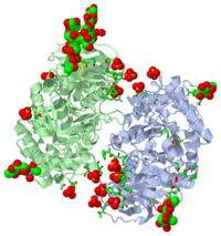
2j25
From Proteopedia
| Line 1: | Line 1: | ||
[[Image:2j25.png|left|200px]] | [[Image:2j25.png|left|200px]] | ||
| - | <!-- | ||
| - | The line below this paragraph, containing "STRUCTURE_2j25", creates the "Structure Box" on the page. | ||
| - | You may change the PDB parameter (which sets the PDB file loaded into the applet) | ||
| - | or the SCENE parameter (which sets the initial scene displayed when the page is loaded), | ||
| - | or leave the SCENE parameter empty for the default display. | ||
| - | --> | ||
{{STRUCTURE_2j25| PDB=2j25 | SCENE= }} | {{STRUCTURE_2j25| PDB=2j25 | SCENE= }} | ||
===PARTIALLY DEGLYCOSYLATED GLUCOCERAMIDASE=== | ===PARTIALLY DEGLYCOSYLATED GLUCOCERAMIDASE=== | ||
| - | |||
| - | <!-- | ||
| - | The line below this paragraph, {{ABSTRACT_PUBMED_17139081}}, adds the Publication Abstract to the page | ||
| - | (as it appears on PubMed at http://www.pubmed.gov), where 17139081 is the PubMed ID number. | ||
| - | --> | ||
{{ABSTRACT_PUBMED_17139081}} | {{ABSTRACT_PUBMED_17139081}} | ||
| Line 24: | Line 13: | ||
*[[Acid-beta-glucosidase|Acid-beta-glucosidase]] | *[[Acid-beta-glucosidase|Acid-beta-glucosidase]] | ||
*[[Partially deglycosylated acid-beta-glucosidase|Partially deglycosylated acid-beta-glucosidase]] | *[[Partially deglycosylated acid-beta-glucosidase|Partially deglycosylated acid-beta-glucosidase]] | ||
| - | *[[Structure Gallery Generator|Structure Gallery Generator]] | ||
*[[Treatment of Gaucher disease|Treatment of Gaucher disease]] | *[[Treatment of Gaucher disease|Treatment of Gaucher disease]] | ||
*[[User:Boris Brumshtein|User:Boris Brumshtein]] | *[[User:Boris Brumshtein|User:Boris Brumshtein]] | ||
Revision as of 17:19, 25 July 2012
Contents |
PARTIALLY DEGLYCOSYLATED GLUCOCERAMIDASE
Gaucher disease is caused by mutations in the gene encoding acid-beta-glucosidase. A recombinant form of this enzyme, Cerezyme, is used to treat Gaucher disease patients by ;enzyme-replacement therapy'. Crystals of Cerezyme after its partial deglycosylation were obtained earlier and the structure was solved to 2.0 A resolution [Dvir et al. (2003), EMBO Rep. 4, 704-709]. The crystal structure of unmodified Cerezyme is now reported, in which a substantial number of sugar residues bound to three asparagines via N-glycosylation could be visualized. The structure of intact fully glycosylated Cerezyme is virtually identical to that of the partially deglycosylated enzyme. However, the three loops at the entrance to the active site, which were previously observed in alternative conformations, display additional variability in their structures. Comparison of the structure of acid-beta-glucosidase with that of xylanase, a bacterial enzyme from a closely related protein family, demonstrates a close correspondence between the active-site residues of the two enzymes.
Structural comparison of differently glycosylated forms of acid-beta-glucosidase, the defective enzyme in Gaucher disease., Brumshtein B, Wormald MR, Silman I, Futerman AH, Sussman JL, Acta Crystallogr D Biol Crystallogr. 2006 Dec;62(Pt 12):1458-65. Epub 2006, Nov 23. PMID:17139081
From MEDLINE®/PubMed®, a database of the U.S. National Library of Medicine.
About this Structure
2j25 is a 2 chain structure of Acid-beta-glucosidase with sequence from Homo sapiens. Full crystallographic information is available from OCA.
See Also
- Acid-beta-glucosidase
- Partially deglycosylated acid-beta-glucosidase
- Treatment of Gaucher disease
- User:Boris Brumshtein
- Velaglucerase alfa
Reference
- Brumshtein B, Wormald MR, Silman I, Futerman AH, Sussman JL. Structural comparison of differently glycosylated forms of acid-beta-glucosidase, the defective enzyme in Gaucher disease. Acta Crystallogr D Biol Crystallogr. 2006 Dec;62(Pt 12):1458-65. Epub 2006, Nov 23. PMID:17139081 doi:S0907444906038303
Categories: Glucosylceramidase | Homo sapiens | Brumshtein, B. | Futerman, A H. | Silman, I. | Sussman, J L. | Wormald, M R. | Alternative initiation | Cerezyme hydrolase | Disease mutation | Gaucher disease | Glucocerebrosidase | Glucosidase | Glycoprotein | Glycosidase | Hydrolase | Lipid metabolism | Lysosome | Membrane | Sphingolipid | Sphingolipid metabolism

