We apologize for Proteopedia being slow to respond. For the past two years, a new implementation of Proteopedia has been being built. Soon, it will replace this 18-year old system. All existing content will be moved to the new system at a date that will be announced here.
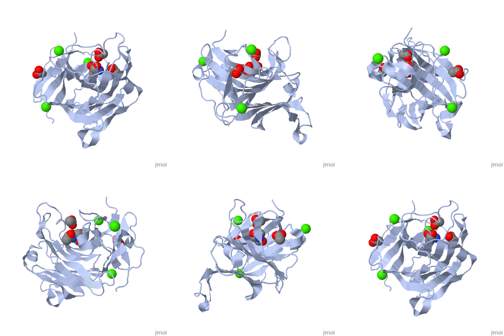
3h0o
From Proteopedia
(Difference between revisions)
| Line 1: | Line 1: | ||
[[Image:3h0o.png|left|200px]] | [[Image:3h0o.png|left|200px]] | ||
| - | <!-- | ||
| - | The line below this paragraph, containing "STRUCTURE_3h0o", creates the "Structure Box" on the page. | ||
| - | You may change the PDB parameter (which sets the PDB file loaded into the applet) | ||
| - | or the SCENE parameter (which sets the initial scene displayed when the page is loaded), | ||
| - | or leave the SCENE parameter empty for the default display. | ||
| - | --> | ||
{{STRUCTURE_3h0o| PDB=3h0o | SCENE= }} | {{STRUCTURE_3h0o| PDB=3h0o | SCENE= }} | ||
===The importance of CH-Pi stacking interactions between carbohydrate and aromatic residues in truncated Fibrobacter succinogenes 1,3-1,4-beta-D-glucanase=== | ===The importance of CH-Pi stacking interactions between carbohydrate and aromatic residues in truncated Fibrobacter succinogenes 1,3-1,4-beta-D-glucanase=== | ||
| - | |||
| - | <!-- | ||
| - | The line below this paragraph, {{ABSTRACT_PUBMED_16246371}}, adds the Publication Abstract to the page | ||
| - | (as it appears on PubMed at http://www.pubmed.gov), where 16246371 is the PubMed ID number. | ||
| - | --> | ||
{{ABSTRACT_PUBMED_16246371}} | {{ABSTRACT_PUBMED_16246371}} | ||
==About this Structure== | ==About this Structure== | ||
| - | [[3h0o]] is a 1 chain structure with sequence from [http://en.wikipedia.org/wiki/Fibrobacter_succinogenes Fibrobacter succinogenes]. Full crystallographic information is available from [http://oca.weizmann.ac.il/oca-bin/ocashort?id=3H0O OCA]. | + | [[3h0o]] is a 1 chain structure of [[Glucanase]] with sequence from [http://en.wikipedia.org/wiki/Fibrobacter_succinogenes Fibrobacter succinogenes]. Full crystallographic information is available from [http://oca.weizmann.ac.il/oca-bin/ocashort?id=3H0O OCA]. |
| + | |||
| + | ==See Also== | ||
| + | *[[Glucanase|Glucanase]] | ||
| + | *[[Molecular Playground/1%2C3-1%2C4-beta-D-glucanase|Molecular Playground/1%2C3-1%2C4-beta-D-glucanase]] | ||
==Reference== | ==Reference== | ||
| - | <ref group="xtra">PMID: | + | <ref group="xtra">PMID:016246371</ref><ref group="xtra">PMID:012842475</ref><references group="xtra"/> |
[[Category: Fibrobacter succinogenes]] | [[Category: Fibrobacter succinogenes]] | ||
[[Category: Licheninase]] | [[Category: Licheninase]] | ||
Revision as of 02:03, 26 July 2012
| |||||||
| 3h0o, resolution 1.40Å () | |||||||
|---|---|---|---|---|---|---|---|
| Ligands: | , , | ||||||
| Activity: | Licheninase, with EC number 3.2.1.73 | ||||||
| Related: | 1mve, 1zm1, 3hr9 | ||||||
| |||||||
| Resources: | FirstGlance, OCA, RCSB, PDBsum | ||||||
| Coordinates: | save as pdb, mmCIF, xml | ||||||
Contents |
The importance of CH-Pi stacking interactions between carbohydrate and aromatic residues in truncated Fibrobacter succinogenes 1,3-1,4-beta-D-glucanase
Template:ABSTRACT PUBMED 16246371
About this Structure
3h0o is a 1 chain structure of Glucanase with sequence from Fibrobacter succinogenes. Full crystallographic information is available from OCA.
See Also
Reference
- Tsai LC, Shyur LF, Cheng YS, Lee SH. Crystal structure of truncated Fibrobacter succinogenes 1,3-1,4-beta-D-glucanase in complex with beta-1,3-1,4-cellotriose. J Mol Biol. 2005 Dec 2;354(3):642-51. Epub 2005 Sep 30. PMID:16246371 doi:http://dx.doi.org/10.1016/j.jmb.2005.09.041
- Tsai LC, Shyur LF, Lee SH, Lin SS, Yuan HS. Crystal structure of a natural circularly permuted jellyroll protein: 1,3-1,4-beta-D-glucanase from Fibrobacter succinogenes. J Mol Biol. 2003 Jul 11;330(3):607-20. PMID:12842475

