We apologize for Proteopedia being slow to respond. For the past two years, a new implementation of Proteopedia has been being built. Soon, it will replace this 18-year old system. All existing content will be moved to the new system at a date that will be announced here.
3h0o
From Proteopedia
(Difference between revisions)
| Line 1: | Line 1: | ||
| - | + | ==The importance of CH-Pi stacking interactions between carbohydrate and aromatic residues in truncated Fibrobacter succinogenes 1,3-1,4-beta-D-glucanase== | |
| - | + | <StructureSection load='3h0o' size='340' side='right' caption='[[3h0o]], [[Resolution|resolution]] 1.40Å' scene=''> | |
| - | + | == Structural highlights == | |
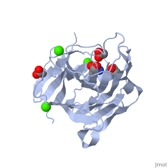
| + | <table><tr><td colspan='2'>[[3h0o]] is a 1 chain structure with sequence from [http://en.wikipedia.org/wiki/Fibrobacter_succinogenes Fibrobacter succinogenes]. Full crystallographic information is available from [http://oca.weizmann.ac.il/oca-bin/ocashort?id=3H0O OCA]. For a <b>guided tour on the structure components</b> use [http://oca.weizmann.ac.il/oca-docs/fgij/fg.htm?mol=3H0O FirstGlance]. <br> | ||
| + | </td></tr><tr id='ligand'><td class="sblockLbl"><b>[[Ligand|Ligands:]]</b></td><td class="sblockDat"><scene name='pdbligand=ACT:ACETATE+ION'>ACT</scene>, <scene name='pdbligand=CA:CALCIUM+ION'>CA</scene>, <scene name='pdbligand=TRS:2-AMINO-2-HYDROXYMETHYL-PROPANE-1,3-DIOL'>TRS</scene></td></tr> | ||
| + | <tr id='related'><td class="sblockLbl"><b>[[Related_structure|Related:]]</b></td><td class="sblockDat">[[1mve|1mve]], [[1zm1|1zm1]], [[3hr9|3hr9]]</td></tr> | ||
| + | <tr id='activity'><td class="sblockLbl"><b>Activity:</b></td><td class="sblockDat"><span class='plainlinks'>[http://en.wikipedia.org/wiki/Licheninase Licheninase], with EC number [http://www.brenda-enzymes.info/php/result_flat.php4?ecno=3.2.1.73 3.2.1.73] </span></td></tr> | ||
| + | <tr id='resources'><td class="sblockLbl"><b>Resources:</b></td><td class="sblockDat"><span class='plainlinks'>[http://oca.weizmann.ac.il/oca-docs/fgij/fg.htm?mol=3h0o FirstGlance], [http://oca.weizmann.ac.il/oca-bin/ocaids?id=3h0o OCA], [http://www.rcsb.org/pdb/explore.do?structureId=3h0o RCSB], [http://www.ebi.ac.uk/pdbsum/3h0o PDBsum]</span></td></tr> | ||
| + | </table> | ||
| + | == Evolutionary Conservation == | ||
| + | [[Image:Consurf_key_small.gif|200px|right]] | ||
| + | Check<jmol> | ||
| + | <jmolCheckbox> | ||
| + | <scriptWhenChecked>select protein; define ~consurf_to_do selected; consurf_initial_scene = true; script "/wiki/ConSurf/h0/3h0o_consurf.spt"</scriptWhenChecked> | ||
| + | <scriptWhenUnchecked>script /wiki/extensions/Proteopedia/spt/initialview01.spt</scriptWhenUnchecked> | ||
| + | <text>to colour the structure by Evolutionary Conservation</text> | ||
| + | </jmolCheckbox> | ||
| + | </jmol>, as determined by [http://consurfdb.tau.ac.il/ ConSurfDB]. You may read the [[Conservation%2C_Evolutionary|explanation]] of the method and the full data available from [http://bental.tau.ac.il/new_ConSurfDB/chain_selection.php?pdb_ID=2ata ConSurf]. | ||
| + | <div style="clear:both"></div> | ||
| + | <div style="background-color:#fffaf0;"> | ||
| + | == Publication Abstract from PubMed == | ||
| + | Fibrobacter succinogenes 1,3-1,4-beta-D-glucanase (Fsbeta-glucanase) catalyzes the specific hydrolysis of beta-1,4 glycosidic bonds adjacent to beta-1,3 linkages in beta-D-glucans or lichenan. This is the first report to elucidate the crystal structure of a truncated Fsbeta-glucanase (TFsbeta-glucanase) in complex with beta-1,3-1,4-cellotriose, a major product of the enzyme reaction. The crystal structures, at a resolution of 2.3 angstroms, reveal that the overall fold of TFsbeta-glucanase remains virtually unchanged upon sugar binding. The enzyme accommodates five glucose residues, forming a concave active cleft. The beta-1,3-1,4-cellotriose with subsites -3 to -1 bound to the active cleft of TFsbeta-glucanase with its reducing end subsite -1 close to the key catalytic residues Glu56 and Glu60. All three subsites of the beta-1,3-1,4-cellotriose adopted a relaxed C(1)4 conformation, with a beta-1,3 glycosidic linkage between subsites -2 and -1, and a beta-1,4 glycosidic linkage between subsites -3 and -2. On the basis of the enzyme-product complex structure observed in this study, a catalytic mechanism and substrate binding conformation of the active site of TFsbeta-glucanase is proposed. | ||
| - | + | Crystal structure of truncated Fibrobacter succinogenes 1,3-1,4-beta-D-glucanase in complex with beta-1,3-1,4-cellotriose.,Tsai LC, Shyur LF, Cheng YS, Lee SH J Mol Biol. 2005 Dec 2;354(3):642-51. Epub 2005 Sep 30. PMID:16246371<ref>PMID:16246371</ref> | |
| - | + | ||
| + | From MEDLINE®/PubMed®, a database of the U.S. National Library of Medicine.<br> | ||
| + | </div> | ||
==See Also== | ==See Also== | ||
*[[Glucanase|Glucanase]] | *[[Glucanase|Glucanase]] | ||
*[[Molecular Playground/1%2C3-1%2C4-beta-D-glucanase|Molecular Playground/1%2C3-1%2C4-beta-D-glucanase]] | *[[Molecular Playground/1%2C3-1%2C4-beta-D-glucanase|Molecular Playground/1%2C3-1%2C4-beta-D-glucanase]] | ||
| - | + | == References == | |
| - | == | + | <references/> |
| - | < | + | __TOC__ |
| + | </StructureSection> | ||
[[Category: Fibrobacter succinogenes]] | [[Category: Fibrobacter succinogenes]] | ||
[[Category: Licheninase]] | [[Category: Licheninase]] | ||
| - | [[Category: Hsiao, C H | + | [[Category: Hsiao, C H]] |
| - | [[Category: Tsai, L C | + | [[Category: Tsai, L C]] |
[[Category: 3-1]] | [[Category: 3-1]] | ||
[[Category: 4-beta-d-glucanase]] | [[Category: 4-beta-d-glucanase]] | ||
Revision as of 06:05, 18 December 2014
The importance of CH-Pi stacking interactions between carbohydrate and aromatic residues in truncated Fibrobacter succinogenes 1,3-1,4-beta-D-glucanase
| |||||||||||