Acid-beta-glucosidase
From Proteopedia
(Difference between revisions)
| Line 19: | Line 19: | ||
*[[Beta glucosidase]]<br /> | *[[Beta glucosidase]]<br /> | ||
*[[Ceramide]]. | *[[Ceramide]]. | ||
| - | |||
| - | Several x-ray structures of the enzyme have been solved. [http://www.pdb.org PDB] codes: | ||
| - | |||
| - | [[1ogs]] - the first structure of acid-beta-glucosidase, the enzyme involved in Gacher disease. Structure of deglycosylated Cerezyme™. <ref>PMID:12792654</ref> | ||
| - | [[2f61]]; [[2nt0]]; [[2nt1]]; [[2v3d]]; [[2v3e]]; [[2v3f]]; [[2nsx]]; [[2j25]] | ||
| - | |||
| - | [[1y7v]] - acid-beta-glucosidase conjugated with an irreversible inhibitor, conduritol-B-epoxide (CBE); | ||
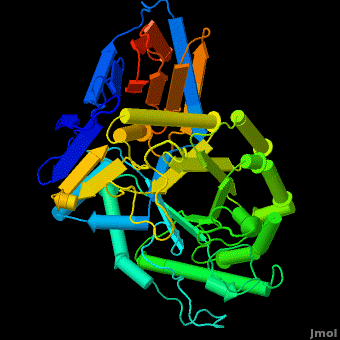
The entrance to the active site of the enzyme is confined by three loops (Loop-1, residues 346-349; Loop-2, 393-399; Loop-3, 312-319), which have been observed in a number of conformations. Loop-1 is found to be involved in crystal contact interactions between the two individual protein molecules in crystals. Its conformation was not found to change significantly in various crystals of the enzyme. In contrast, loops-2 and 3 were detected in several different stable conformations, and displayed varying conformations in different structures even in the absence of an inhibitor in the active site. These loops were found to change the shape of the entrance as well as several properties of the active site. | The entrance to the active site of the enzyme is confined by three loops (Loop-1, residues 346-349; Loop-2, 393-399; Loop-3, 312-319), which have been observed in a number of conformations. Loop-1 is found to be involved in crystal contact interactions between the two individual protein molecules in crystals. Its conformation was not found to change significantly in various crystals of the enzyme. In contrast, loops-2 and 3 were detected in several different stable conformations, and displayed varying conformations in different structures even in the absence of an inhibitor in the active site. These loops were found to change the shape of the entrance as well as several properties of the active site. | ||
Revision as of 10:29, 8 January 2023
| |||||||||||
References
- ↑ Grabowski GA. Gaucher disease and other storage disorders. Hematology Am Soc Hematol Educ Program. 2012;2012:13-8. doi:, 10.1182/asheducation-2012.1.13. PMID:23233555 doi:http://dx.doi.org/10.1182/asheducation-2012.1.13
Additional Resources
Metabolic Disorders
Carbohydrate Metabolism
Treatment of Gaucher disease
Proteopedia Page Contributors and Editors (what is this?)
Michal Harel, Boris Brumshtein, Alexander Berchansky, Joel L. Sussman, Eran Hodis, David Canner
