3h0o
From Proteopedia
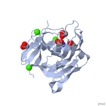
| |||||||
| 3h0o, resolution 1.40Å () | |||||||
|---|---|---|---|---|---|---|---|
| Ligands: | , , | ||||||
| Activity: | Licheninase, with EC number 3.2.1.73 | ||||||
| Related: | 1mve, 1zm1, 3hr9 | ||||||
| |||||||
| Resources: | FirstGlance, OCA, RCSB, PDBsum | ||||||
| Coordinates: | save as pdb, mmCIF, xml | ||||||
Contents |
The importance of CH-Pi stacking interactions between carbohydrate and aromatic residues in truncated Fibrobacter succinogenes 1,3-1,4-beta-D-glucanase
Template:ABSTRACT PUBMED 16246371
About this Structure
3h0o is a 1 chain structure with sequence from Fibrobacter succinogenes. Full crystallographic information is available from OCA.
See Also
Reference
- Tsai LC, Shyur LF, Cheng YS, Lee SH. Crystal structure of truncated Fibrobacter succinogenes 1,3-1,4-beta-D-glucanase in complex with beta-1,3-1,4-cellotriose. J Mol Biol. 2005 Dec 2;354(3):642-51. Epub 2005 Sep 30. PMID:16246371 doi:http://dx.doi.org/10.1016/j.jmb.2005.09.041
- Tsai LC, Shyur LF, Lee SH, Lin SS, Yuan HS. Crystal structure of a natural circularly permuted jellyroll protein: 1,3-1,4-beta-D-glucanase from Fibrobacter succinogenes. J Mol Biol. 2003 Jul 11;330(3):607-20. PMID:12842475
